Translate this page into:
Primary B-cell Non-Hodgkin Lymphoma of Gallbladder Presenting as Cholecystitis
Address for correspondence: Dr. Richa Katiyar, E-mail: drrichakati@gmail.com
This is an open-access article distributed under the terms of the Creative Commons Attribution-Noncommercial-Share Alike 3.0 Unported, which permits unrestricted use, distribution, and reproduction in any medium, provided the original work is properly cited.
This article was originally published by Medknow Publications & Media Pvt Ltd and was migrated to Scientific Scholar after the change of Publisher.
Sir,
Primary gallbladder lymphoma (PGBL) is defined as an extranodal lymphoma arising and confined to gallbladder with/without contiguous lymph node involvement and distant spread.[1] Less than 50 cases of PGBL have been reported till 2010.[2]
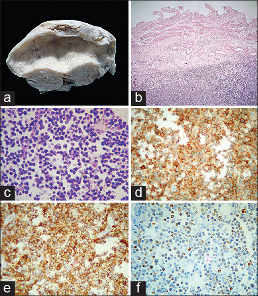
A 48-year-old woman presented with malaise and sudden onset of abdominal pain. Her blood pressure was 118/80 mm Hg. Tenderness was present in the right upper quadrant of the abdomen. Laboratory tests revealed reactive HBsAg and serum creatinine 2.5 mg/dl, while other investigations were normal. Abdominal ultrasound showed irregular edematous gallbladder wall, multiple calculi and sludge particles suggestive of cholelithiasis, acute cholecystitis and empyema gallbladder. Sizeable retroperitoneal lymphadenopathy was absent. Patient underwent cholecystectomy elsewhere. Specimen was submitted to author AA. Grossly, gallbladder was 10 cm × 4 cm with multiple small yellow-brown multifaceted stones and normal velvety mucosa. Gallbladder wall was thickened (1.5–2 cm) with rubbery and gray white cut surface [Figure 1a]. Microscopically, intact gallbladder mucosa was infiltrated by lymphocytes and plasma cells. However, muscular and adventitial layers showed diffuse infiltrates of medium-sized atypical lymphoid cells with centrocyte and centroblast-like morphology [Figure 1b and c]. On immunohistochemistry, CD45 and CD20 positive B-cells expressing scattered positivity for bcl-2 [Figure 1d–f] and negative expression for CD3, CD5, CD15, CD23, CD30 and cyclin D1 were seen. A diagnosis of non-Hodgkin lymphoma (NHL) B-cell diffuse type, not further specified was made. Patient died on postoperative 2nd day.

- (a) Gross photograph showing nodular thickening of gallbladder wall and unremarkable mucosa. (b) Diffuse infiltrates of lymphoid cells in the adventitia of gall bladder with sparing of mucosa and muscularis (H and E, ×40). (c) Relatively uniform medium-sized atypical lymphoid cells on a background of lymphoglandular bodies (H and E, ×400). (d) Diffuse strong membranous staining of atypical lymphoid cells (CD45, ×400). (e) Diffuse strong membranous staining of atypical lymphoid cells (CD20, ×400). (f) Scattered nuclear staining of atypical lymphoid cells (bcl-2, ×100)
This is an extremely rare occurrence of PGBL which was clinically diagnosed as cholecystitis, cholelithiasis and empyema of gallbladder. Most PGBLs clinically present with symptoms of cholecystitis.[2] Similarly, a clinically diagnosed acute cholecystitis with empyema turned out to be B-cell NHL of gallbladder.[3] Submucosal homogenous wall thickening of gallbladder in radiology correlate with pathological diagnosis of lymphoma.[4] For definite diagnosis, histopathology and immunohistochemistry are mandatory. Treatment options for PGBL include surgery with the use of chemotherapy in disseminated disease and inoperable cases.
ACKNOWLEDGMENT
Authors are sincerely thankful to Dr. Sanjay Navani of Mumbai for doing immunohistochemistry in this case.
REFERENCES
- World Health Organization Classification of Tumours: Pathology and Genetics of Tumours of the Digestive System. Lyon: IARC Press; 2000.
- [Google Scholar]
- Gall bladder and extrahepatic bile duct lymphomas: Clinicopathological observations and biological implications. Am J Surg Pathol. 2010;34:1277-86.
- [Google Scholar]
- Primary malignant lymphoma of the gallbladder: A case report and literature review. Br J Radiol. 2009;82:e15-9.
- [Google Scholar]


