Translate this page into:
An Incidental Primary Papillary Carcinoma Arising in a Thyroglossal Duct Cyst: Report of a Rare Finding
Address for correspondence: Dr. Sujata Jetley, E-mail: sujatajetley@gmail.com
This is an open access article distributed under the terms of the Creative Commons Attribution NonCommercial ShareAlike 3.0 License, which allows others to remix, tweak, and build upon the work non commercially, as long as the author is credited and the new creations are licensed under the identical terms.
This article was originally published by Medknow Publications & Media Pvt Ltd and was migrated to Scientific Scholar after the change of Publisher.
Abstract
The thyroglossal duct cysts (TGDCs) are the most common congenital anomaly of the thyroid, usually manifested as painless midline neck mass. Malignancy is very rare and is reported in around 1% of cases as an incidental finding after histopathological evaluation of resected cyst. Papillary carcinoma is the most common carcinoma reported in TGDC. Here, we report a case of 17-year-old-female, who presented with a gradually increasing midline neck mass which moves with swallowing. On imaging a diagnosis of infected TGDC was made. The Sistrunk operation was done and a diagnosis of primary papillary carcinoma arising in a TGDC was rendered histopathologically. The contemporary appearance of papillary carcinoma thyroid was reported in about 20% cases of TGDC carcinoma, thus it is essential to differentiate primary papillary carcinoma arising in a TGDC from those of metastatic papillary carcinoma thyroid by strict diagnostic criteria.
Keywords
Papillary carcinoma
Sistrunk operation
thyroglossal duct
INTRODUCTION
Thyroglossal tract is an epithelial tract which is formed as a result of descent of thyroid gland from foramen cecum to its normal location just below the thyroid cartilage and this tract usually disappears between 5th and 10th gestational weeks. The thyroglossal duct cyst (TGDC) is formed either as a result of incomplete atrophy of thyroglossal tract, or retained epithelial cyst. The remnants of the thyroglossal tract may be present as “cysts, tract or duct, a fistula or as ectopic thyroid within a cyst or duct.”[12] TGDC accounts for about 70% of all midline neck masses seen in children. However, it is responsible for only 7% cases in adults.[3]
Primary malignancy in TGDC is extremely rare and among all cases of TGDC, carcinoma is reported only in 0.7–1% of cases.[45] Papillary carcinoma (80–85%) is the most common carcinoma reported in TGDC, followed by squamous cell carcinoma (5–6%).[6]
Here, we report a case of 17-year-old-female presented with a midline neck mass, which was diagnosed and operated for infected TGDC. However, on final histopathology, a diagnosis of papillary carcinoma arising in a TGDC was made.
CASE REPORT
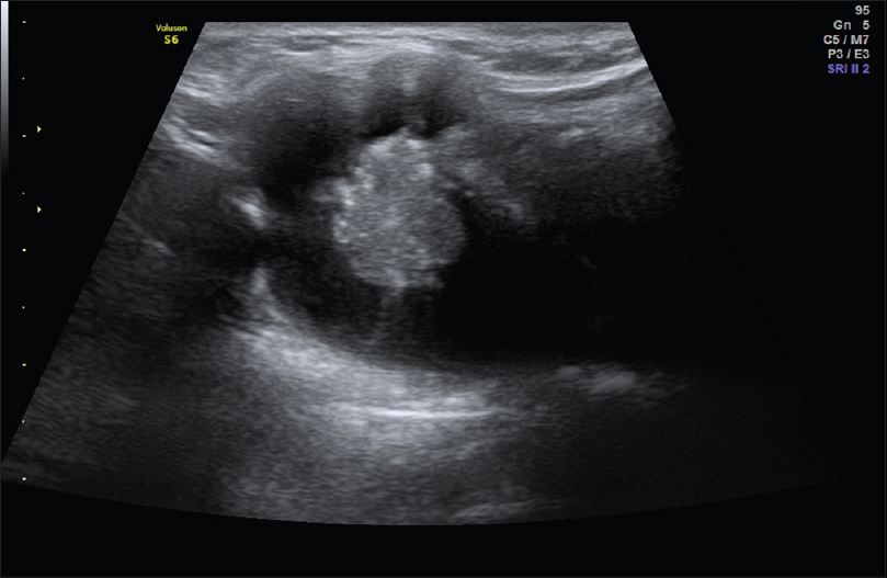
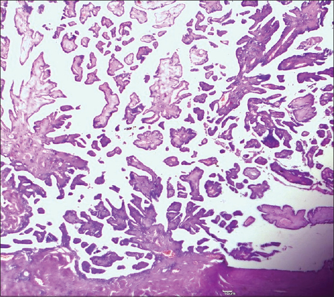
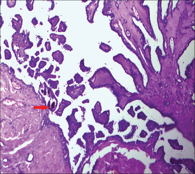
A 17-year-old-female patient presented to Otorhinolaryngology Outpatient Department with a history of swelling over the anterior aspect of neck with a gradual increase in size for last 6 months. There was no history of pain, dysphagia, or hoarseness of voice. There was no history of cold or hot intolerance, tremor, palpitation, diarrhea, constipation, any menstrual irregularities, or any radiation exposure. On examination, a single, well-defined, painless, soft to firm swelling was seen in the midline of neck, measuring about 2 cm × 1.5 cm. The swelling moved with both swallowings as well as the movement of tongue. The overlying skin was normal, and there was no palpable cervical lymphnodes. The routine laboratory investigations including complete blood count, liver function test, and kidney function test was within normal range. The thyroid function test was within normal limit. The ultrasonography (USG) of neck revealed a midline complex cystic mass (2 cm × 1.5 cm) above the thyroid isthmus with multiple internal septations and a central large nodule with heterogeneous echogenicity and coarse calcification [Figure 1]. Both lobes of thyroid were normal in size and echo pattern with smooth margins. The isthmus was also normal, and there was no evidence of necrosis or hemorrhage. Thus, a diagnosis of infected TGDC was made. Following this diagnosis, Sistrunk operation was done which consist of resection of the cyst along with a central portion of the hyoid bone and dissection of the thyroglossal tract up to the base of the tongue. The resected specimen sent for histopathological examination. On gross examination, a globular soft tissue mass measuring 2 cm × 1.5 cm was received along with 1.5 cm elongated fibrous tissue attached to globular mass and a piece of bone. On cut section, a unilocular cyst (1 cm × 0.8 cm) filled with brownish material was seen. Microscopic examination showed a cyst lined by cuboidal epithelial cells with foci of papillary projections with broad fibrovascular core [Figure 2]. The papillae were lined by cuboidal epithelial cells, some of which show nuclear clearing and grooves along with the presence of psammoma bodies in the fibrovascular core [Figure 3]. The fibro-collagenous wall of the cyst contains few colloid-filled follicles, blood vessels, and areas of hemorrhage. Thus, a diagnosis of papillary carcinoma arising in a TGDC was rendered histopathologically.

- Ultrasonography showing a well-defined lobulated anechoic lesion with central echogenic nodule

- Microphotograph showing papillary projections arising from cyst wall (H and E, ×100)

- Microphotograph showing papillary projections with broad fibrovascular core along with presence of psammoma bodies in the core of papillae (red arrow) (H and E, ×400)
The patient is in regular follow-up and remains asymptomatic for last 1 year. There were no signs of any recurrence. Repeated USG of neck showed normal thyroid scan.
DISCUSSION
TGDCs are the most common congenital anomaly of the thyroid, usually manifested as enlarging, painless midline neck mass. This is the most common cause of midline neck mass in children (75%), but this is reported only in 7% of adult population. In the majority of the cases, TGDCs are benign. However, malignancy is reported in around 1% of cases and is usually diagnosed as an incidental finding after histopathological evaluation of resected TGDC.[67] Thyrogenic carcinoma is the most common malignant tumor of TGDC, which originates from thyroglossal tract remnant followed by squamous cell carcinoma which originates from metaplastic columnar cells of the thyroglossal duct lining.[1]
Histologically papillary carcinoma (80–85%) is the most common carcinoma arising in a TGDC. Except for medullary carcinoma thyroid which originates from parafollicular cells, other thyroid tumors such as follicular, mixed papillay-follicular and Hurthle cell carcinoma have also been reported, though infrequently.[8] Although seen in only about 5% of cases, squamous cell carcinoma of TGDC is considered as only primary TGDC tumor by some authors, because other malignancy usually develop in ectopic thyroid tissue.[9] An asymptomatic midline soft to firm, painless swelling at the anterior aspect of neck, which moves with deglutition is the usual presentation of TGDC. The possibility of TGDC carcinoma should be suspected if the cyst is hard, fixed or irregular.[10] Secondary, infection or formation of cutaneous fistula is a common manifestation of TGDC.[6]
The contemporary appearance of papillary carcinoma thyroid was reported in about 20% cases of TGDC carcinoma,[11] thus it is essential to differentiate primary papillary carcinoma arising in a TGDC from those of metastatic papillary carcinoma thyroid. The following three criteria are essential for the diagnosis of primary carcinoma arising in a TGDC. (1) Presence of carcinomatous component in the wall of TGDC, (2) presence of normal thyroid tissue adjacent to the tumor, (3) presence of clinically normal thyroid gland without evidence of any primary thyroid carcinoma.[1213] Our case fulfills all these three criteria. Sistrunk operation is the procedure of choice for this condition,[11] however, some authors recommended additional total thyroidectomy, if the carcinomatous component is more than 1 cm in size and even if the tumor is <1 cm but show enlarged cervical lymph node, tumor invasion of the cyst wall or abnormal thyroid gland.[14] Although prognosis of papillary carcinoma arising in a TGDC is excellent with reported occurrence of metastatic lesion in <2% of cases,[5] however, considering the multifocal nature of papillary carcinoma, a careful long-term follow-up is recommended.
Financial support and sponsorship
Nil.
Conflicts of interest
There are no conflicts of interest.
REFERENCES
- Thyroglossal duct carcinoma in children: Case presentation and review of the literature. Thyroid. 2004;14:777-85.
- [Google Scholar]
- Diagnosis of papillary carcinoma in a thyroglossal duct cyst by fine-needle aspiration biopsy. Arch Pathol Lab Med. 2000;124:139-42.
- [Google Scholar]
- Papillary carcinoma arising in thyroglossal duct cyst in the lateral neck. Pathol Res Pract. 2013;209:674-8.
- [Google Scholar]
- Papillary carcinoma in thyroglossal duct cyst: An unusual case. Egypt J Ear Nose Throat Allied Sci. 2014;15:45-7.
- [Google Scholar]
- Papillary carcinoma arising from thyroglossal duct cyst with thyroid and lateral neck metastasis. Int J Surg Case Rep. 2013;4:704-7.
- [Google Scholar]
- Squamous cell carcinoma arising in a thyroglossal duct cyst. Am Surg. 1974;40:290-4.
- [Google Scholar]
- Management of well-differentiated thyroid carcinoma presenting within a thyroglossal duct cyst. J Surg Oncol. 2002;79:134-9.
- [Google Scholar]
- Adenocarcinoma originating in the thyroglossal duct. Ann Otol Rhinol Laryngol. 1976;85(2 pt 1):286-90.
- [Google Scholar]
- Thyroglossal duct cyst carcinoma: Case series. J Otolaryngol Head Neck Surg. 2011;40:151-6.
- [Google Scholar]


