
Translate this page into:
Comparative evaluation of MIC values of Trichosporon spp. by MTT assay and CLSI M27-A3 broth microdilution reference methods

*Corresponding author: Thayanidhi Premamalini, Department of Microbiology, Sri Ramachandra Medical College and Research Institute, Chennai, Tamil Nadu, India.drtpremamalini@gmail.com
-
Received: ,
Accepted: ,
How to cite this article: Premamalini T, Pillai S, Subramanian A, Kindo AJ. Comparative evaluation of MIC values of Trichosporon spp. by MTT assay and CLSI M27-A3 broth microdilution reference methods. J Lab Physicians. 2024;16:461-5. doi: 10.25259/JLP_33_2024
Abstract
Objectives:
The objective of this study was to determine and compare the minimum inhibitory concentration (MIC) values of Trichosporon spp. by MTT (3-(4, 5-dimethyl-2-thiazolyl)-2, 5-diphenyl- 2H-tetrazoliumbromide) assay, and Clinical and Laboratory Standards Institute M27-3rd edition (CLSI M27-A3) broth microdilution methods.
Materials and Methods:
Antifungal susceptibility testing was done by CLSI M27-A3 broth microdilution and MTT assay for all the 72 Trichosporon isolates after genus specific and Trichosporon asahii specific polymerase chain reaction (PCR). Candida krusei ATCC 6258 was used as the reference strain.
Statistical analysis:
All statistical data were analyzed using the Statistical Package for the Social Sciences, version 17 for Microsoft Windows. The percentage of agreement was calculated using the Type C intraclass correlation coefficient.
Results:
The MICs by MTT assay strongly correlated with those obtained by CLSI M27-A3 method, by being either the same or within 1 dilution of MIC by CLSI method. Furthermore, the ranges of MICs obtained by MTT and CLSI method were all identical in our study. The overall agreement between the two methods for the Trichosporon isolates was good, that is, 90.8% in our study.
Conclusions:
MTT assay can be an alternative method that assists reading of MICs visually with a colored end point, making it easier compared to CLSI M27-A3 method. MTT assay can also be standardized for other yeasts and molds so that antifungal susceptibility tests can be done for different fungi.
Keywords
Trichosporon spp
MTT assay
Clinical and Laboratory Standards Institute M27- 3rd edition
Antifungal susceptibility testing
Colorimetric assay

INTRODUCTION
Trichosporon species are yeast-like fungi that are ubiquitous in nature and found as commensal flora on the human skin, gastrointestinal tract, and mucosal surfaces. It can cause invasive infections, especially among immunocompromised hosts.[1] Many times, this genus gets misidentified as Candida species, and patients are started on fluconazole or echinocandins. However, they have limited or no action against fluconazole or amphotericin-B and are also intrinsically resistant to echinocandins. Hence, performing antifungal susceptibility testing is essential to know the exact sensitivity pattern of a particular isolate and it also helps in reducing mortality as well as prevalence of resistance.[2,3] The Clinical and Laboratory Standards Institute (CLSI) has developed the CLSI M27-A3 document, which is the broth microdilution method for the antifungal susceptibility testing of Trichosporon species and similar protocols for other yeasts and molds. These are widely used in most laboratories.[4] The disadvantage of this method is the observer bias which can happen while performing turbidity measurements.[5]
MTT (3-(4, 5-dimethyl-2-thiazolyl)-2, 5-diphenyl-2Htetrazoliumbromide) assay is a colorimetric assay which quantifies mitochondrial respiration by viable fungi through the reduction of MTT to formazan (violet blue water insoluble molecule) indicating cell metabolic activity. Using this principle, this assay can also help in antifungal susceptibility testing.[6,7] Presumably, due to its positive charge and lipophilic structure, MTT reagent is able to penetrate the cell membrane as well as the inner membrane of mitochondrial cells and is reduced by metabolically active cells.[7] The ease with which minimum inhibitory concentration (MIC) breakpoints can be determined and the potential for automation of colorimetric methods make this method appealing, especially in clinical microbiology laboratories with a larger number of samples.[8]
MATERIALS AND METHODS
The reference strains used as quality control for the susceptibility testing were Candida krusei ATCC 6258. A total of 72 isolates were tested according to the CLSI M27- A3 document, out of which 43 (59.7%) isolates were from urine, 12 (16.7%) isolates were from blood, and 7 (9.7%) isolates grew from samples collected immediately after insertion of a percutaneous nephrostomy tube. Out of the 5 (6.9%) respiratory isolates, three were from sputum, and two were from bronchoalveolar lavage. Four (5.6%) were isolated from pus, and 1 isolate (1.4%) grew from peritoneal dialysate fluid collected from the dialysis bag of a patient who underwent peritoneal dialysis. The strains were characterized phenotypically by cultural characteristics (dry yeast-like colonies), biochemical reaction (urea hydrolysis), and microscopic appearance (arthroconidia, blastoconidia, and pseudohyphae). These were provisionally identified as Trichosporon species, and further, confirmation was done through Trichosporon genus specific polymerase chain reaction (PCR) followed by Trichosporon asahii specific PCR. The seven antifungal drugs along with the range of concentration tested were as follows: Amphotericin B (A9528-50MG, Sigma-Aldrich, USA) (range 0.025–16 μg/mL), fluconazole (32103-25MG, Sigma-Aldrich, USA) (range 0.125–64 μg/mL), itraconazole (32103-25MG, Sigma-Aldrich, USA) (range 0.025–16 μg/mL), voriconazole (PZ0005-5MG, Sigma-Aldrich, USA) (range 0.025–16 μg/mL), posaconazole (32103-5MG, Sigma-Aldrich, USA) (range 0.025–16 μg/mL), ravuconazole (SML1216-5MG, Sigma-Aldrich, USA) (range 0.025–16 μg/mL), and caspofungin diacetate (SML0425-5MG, Sigma-Aldrich) (range 0.015–8 μg/mL). A growth control (inoculum without drug) and drug control (drug without inoculum) were included for each isolate tested. Fresh cultures (24–72 h old) grown on Sabouraud dextrose agar (SDA) were used. The plates were incubated at 35°C, and the results were read after 24 h.
For the MTT assay, the initial steps of the susceptibility plate preparation were according to the micro broth dilution method of CLSI M27-A3 guidelines. Four hours before the endpoint reading (i.e., after 20 h of incubation), the plates were agitated, and 50 μL of working solution of MTT was added to each well and incubated at 37°C for 4 h. The MTT solution was then aspirated, and 100 μL of dimethyl sulfoxide was added to each well to solubilize the insoluble end product formazan. The lowest concentration of the antifungal agent where no color change was visually observed was taken as the MIC in the MTT assay.
The Statistical Package for Social Sciences version 17 for Microsoft windows was used for the analysis of all statistical data. Percentage of agreement was calculated using Type C intraclass correlation coefficient.
RESULTS
The 72 isolates were phenotypically characterized by their dry yeast, such as colonies on the culture plate, microscopic features like arthroconidia, blastoconidia, and pseudohyphae, and hydrolysis of Christensen urea agar. These were provisionally identified as belonging to the genus Trichosporon, and further confirmation was done by Trichosporon genus-specific PCR. Out of the 72 isolates, 65 were identified as T. asahii by T. asahii specific PCR. Antifungal susceptibility tests revealed that the MICs by MTT assay (no color change visually) Figure 1 strongly correlated with those obtained by CLSI M27-A3 method by being either the same or within 1 dilution of MIC by CLSI method. Higher mean MIC was noted for amphotericin-B, fluconazole, and itraconazole for both methods. The mean MIC obtained by CLSI and MTT was 4.68 μg/mL and 6.2 μg/mL for amphotericin-B, 3.8 μg/mL and 4.68 μg/mL for fluconazole, and 1.49 μg/mL and 1.95 μg/mL for itraconazole, respectively. Voriconazole and posaconazole had lower mean MIC in our study with 0.21 μg/mL and 0.23 μg/mL for voriconazole and 0.42 μg/mL and 0.49 μg/mL for posaconazole by CLSI and MTT assays. Ravuconazole had the lowest mean MIC with 0.14 μg/mL and 0.16 μg/mL by CLSI and MTT assay, respectively. Caspofungin, which is intrinsically resistant, had an identical mean MIC of 7.39 μg/mL by both methods. Furthermore, the ranges of MICs obtained by MTT and CLSI methods were all identical in our study [Table 1].
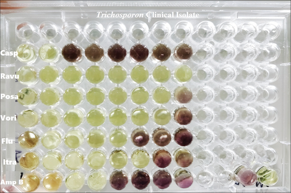
- Antifungal susceptibility testing – MTT (3-(4, 5-dimethyl-2-thiazolyl)-2, 5-diphenyl- 2H-tetrazoliumbromide) assay. Casp: Caspofungin, Ravu: Ravuconazole, Posa: Posaconazole, Vori: Voriconazole, Flu: Fluconazole, Itra: Itraconazole, Amp B: Amphotericin B. Reddish pink wells indicate turbidity with viable fungi. Rest of the wells are not turbid with the absence of viable fungi.
| Antifungal agent | MIC μg/mL | Percentage of Agreement* | |||||
|---|---|---|---|---|---|---|---|
| CLSI | MTT | ||||||
| Mean | ±SD | Range | Mean | ±SD | Range | ||
| Amphotericin B | 4.68 | 2.44 | (2–16) | 6.2 | 2.84 | (2–16) | 70.8 |
| Fluconazole | 3.8 | 10.61 | (0.5–64) | 4.68 | 10.52 | (0.5–64) | 99.7 |
| Itraconazole | 1.49 | 1.94 | (0.25–16) | 1.95 | 2.13 | (0.25–16) | 95.9 |
| Voriconazole | 0.21 | 0.15 | (<0.125–0.5) | 0.23 | 0.15 | (<0.125–0.5) | 97.4 |
| Posaconazole | 0.42 | 0.39 | (<0.125–2) | 0.49 | 0.42 | (0.125–2) | 95.6 |
| Ravuconazole | 0.14 | 0.06 | (<0.125–0.5) | 0.16 | 0.09 | (<0.125–0.5) | 74.5 |
| Caspofungin | 7.39 | 0.45 | (4–>8) | 7.39 | 1.45 | (4–>8) | 100 |
The percentage of agreement was high for caspofungin (100%), fluconazole (99.7%), voriconazole (97.4%), itraconazole (95.9%), and posaconazole (95.6%). The percentage of agreement was little low for ravuconazole (74.5%) and amphotericin B (70.8%). However, the overall agreement between the two methods CLSI and MTT assay for the Trichosporon isolates was good, that is, 90.8% in our study.
DISCUSSION
Antifungal susceptibility testing of Trichosporon species is important since different species can have varied susceptibility patterns. Studies have shown that T. asahii is more resistant to amphotericin-B than triazole compounds, whereas the reverse was seen for the other species in this genus.[9] In other studies, it was reported that all the species in the genus Trichosporon had higher MIC for amphotericin-B and showed poor clinical outcomes with neutropenic patients, indicating that this might not be the drug of choice to treat Trichosporon species infections.[10,11] Trichosporon species also show an intrinsic resistance to echinocandins and poor susceptibility to polyenes.[12] In the study of Paphitou et al., it was suggested that azoles were more potent compared to amphotericin-B for Trichosporon infections and that the fungicidal effect was better for posaconazole, ravuconazole and voriconazole.[10] Since 2010, there have been cases of Trichosporon strains being resistant to voriconazole, and a study in Greece revealed that 38% of Trichosporon isolates showed MIC ≥ 2mg/L for voriconazole.[13] Colorimetric methods of antifungal susceptibility testing are becoming more popular due to the limitation of the CLSI broth microdilution method in terms of turbidity measurement, which is subjective and prone to inter-rater variability.[14] Tetrazolium salts have previously been used to determine the metabolic rates of higher eukaryotic cells and to assess the effects of cytotoxic agents,[15] to provide a non-destructive and continuous spectrophotometric measurement of cell respiration,[16] and to describe differences between the susceptibilities of adherent and non-adherent cells of Candida species.[17] In recent years, there have also been studies where MTT assay was used to check the effectiveness of newer drugs against fungi like Candida albicans.[18]
In our study, higher mean MICs for caspofungin, amphotericin B, fluconazole, and itraconazole were observed on susceptibility testing by MTT assay (endpoint reading- no color change visually) and the overall agreement between CLSI method and MTT assay for antifungal susceptibility testing was 90.8%. In another study by Di Bonaventura et al., 93.7% of agreement was demonstrated between the CLSI method and spectrophotometric evaluation by XTT assay for antifungal susceptibility testing.[19] CLSI-M27 A3 broth microdilution method has been accepted as a standard method for antifungal susceptibility testing of yeasts,and in vitro, determination of MIC values also correlates with the clinical outcome; however, the method is tedious and expensive. The test is affected by the concentration of the inoculum, composition, pH of the medium, and temperature and time for incubation. The reading of MIC is also not easy since observer bias can occur during turbidity measurement.[4] In these situations, a colorimetric method can be used instead, as the interpretation of MIC is more accurate and consistent and can be done even in laboratories with less extensive facilities.
Since comparable results have been obtained by similar studies (90.8% in our study and 93.7% in Di Bonaventura et al.’s study), it can be understood that colorimetric assays can be used as an alternative to the most widely accepted broth microdilution method with regard to antifungal susceptibility testing.[19]
CONCLUSIONS
Trichospon spp. has varied susceptibility patterns and increasing resistance to different antifungal drugs making it imperative to do antifungal susceptibility tests so that appropriate drugs can be started for the patient for a better prognosis. The broth microdilution method according to M27-A3 document guidelines is the standard method for the determination of MICs, although some difficulty may still be encountered in determining the MICs of certain antifungal agents like azoles (trailing effect). A common disadvantage is the subjective differences in MIC interpretation while measuring turbidity.
MTT assay can be an alternative method that assists reading of MICs visually with a colored end point, making the reading of MICs easier, compared to CLSI M27-A3 method. In our study, we have standardized MTT assay for Trichosporon spp., and it can also be standardized for other yeasts and molds so that antifungal susceptibility tests can be done for different fungi.
Acknowledgment
We extend our heartfelt thanks to our institution for providing excellent infrastructures and facilities for research.
Ethical approval
The research/study approved by the Institutional Review Board at Sri Ramachandra Medical College & Research Institute, SRIHER, number IEC-NI/12/MAR/27/14, dated 06th June 2012.
Declaration of patient consent
Patient’s consent was not required as there are no patients in this study.
Conflicts of interest
There are no conflicts of interest.
Use of artificial intelligence (AI)-assisted technology for manuscript preparation
The authors confirm that there was no use of artificial intelligence (AI)-assisted technology for assisting in the writing or editing of the manuscript and no images were manipulated using AI.
Financial support and sponsorship
Nil.
References
- Invasive trichosporonosis caused by Trichosporon asahii and other unusual Trichosporon species at a medical center in Taiwan. Clin Infect Dis. 2009;49:e11-7.
- [CrossRef] [Google Scholar]
- Trichosporon asahii a non-Candida yeast that caused fatal septic shock in a patient without cancer or neutropenia. Clin Infect Dis. 2001;33:E28-30.
- [CrossRef] [Google Scholar]
- Strain typing of Trichosporon asahii clinical isolates by random amplification of polymorphic DNA (RAPD) analysis. J Lab Physicians. 2021;13:245-51.
- [CrossRef] [Google Scholar]
- Comparison of the sensititre yeastone antifungal method with the CLSI M27-A3 reference method to determine the activity of antifungal agents against clinical isolates of Candida spp. Turk J Med Sci. 2020;50:2024-31.
- [CrossRef] [Google Scholar]
- Factors influencing susceptibility testing of antifungal drugs: A critical review of document M27-A4 from the Clinical and Laboratory Standards Institute (CLSI) Braz J Microbiol Publ Braz Soc Microbiol. 2020;51:1791-800.
- [CrossRef] [Google Scholar]
- Comparison of a photometric method with standardized methods of antifungal susceptibility testing of yeasts. J Clin Microbiol. 1997;35:2878-82.
- [CrossRef] [Google Scholar]
- The MTT assay: Utility, limitations, pitfalls, and interpretation in bulk and single-cell analysis. Int J Mol Sci. 2021;22:12827.
- [CrossRef] [Google Scholar]
- Evaluation of a novel colorimetric broth microdilution method for antifungal susceptibility testing of yeast isolates. J Clin Microbiol. 1994;32:1992-6.
- [CrossRef] [Google Scholar]
- Susceptibility patterns and molecular identification of Trichosporon. Antimicrob Agents Chemother. 2005;49:4026-34.
- [CrossRef] [Google Scholar]
- In vitro antifungal susceptibilities of Trichosporon species. Antimicrob Agents Chemother. 2002;46:1144-6.
- [CrossRef] [Google Scholar]
- Trichosporon beigelii an emerging pathogen resistant to amphotericin B. J Clin Microbiol. 1990;28:1616-22.
- [CrossRef] [Google Scholar]
- The epidemiology, genotypes, antifungal susceptibility of Trichosporon species, and the impact of voriconazole on Trichosporon fungemia patients. J Formos Med Assoc. 2021;120:1686-94.
- [CrossRef] [Google Scholar]
- Emerging pan-resistance in Trichosporon species: A case report. BMC Infect Dis. 2016;16:148.
- [CrossRef] [Google Scholar]
- Development and validation of a colorimetric antifungal susceptibility testing method for the dimorphic fungus Talaromyces marneffei. Med Mycol. 2023;61:myad111.
- [CrossRef] [Google Scholar]
- Rapid colorimetric assay for cellular growth and survival: Application to proliferation and cytotoxicity assays. J Immunol Methods. 1983;65:55-63.
- [CrossRef] [Google Scholar]
- Nondestructive and continuous spectrophotometric measurement of cell respiration using a tetrazolium-formazan microemulsion. J Microbiol Methods. 1995;22:283-92.
- [CrossRef] [Google Scholar]
- Comparisons of the susceptibilities of planktonic and adherent Candida albicans to antifungal agents: A modified XTT tetrazolium assay using synchronised C. albicans cells. J Med Vet. 1996;34:149-52.
- [CrossRef] [Google Scholar]
- Antifungal properties of hydrazine-based compounds against Candida albicans. Antibiot Basel Switz. 2023;12:1043.
- [CrossRef] [Google Scholar]
- Biofilm formation by the emerging fungal pathogen Trichosporon asahii Development, architecture, and antifungal resistance. Antimicrob Agents Chemother. 2006;50:3269-76.
- [CrossRef] [Google Scholar]


