
Translate this page into:
Biofilm colonization in chronic treatment refractory infections presenting with discharging sinuses: A study in a tertiary care hospital of Eastern India
Address for correspondence: Dr. Hirak Jyoti Raj, J-5, Sahapore Goverment Housing Estate, New Alipore, Kolkata - 700 038, West Bengal, India. E-mail: hirak17raj@gmail.com
-
Received: ,
Accepted: ,
This is an open access article distributed under the terms of the Creative Commons Attribution-NonCommercial-ShareAlike 3.0 License, which allows others to remix, tweak, and build upon the work non-commercially, as long as the author is credited and the new creations are licensed under the identical terms.
This article was originally published by Medknow Publications & Media Pvt Ltd and was migrated to Scientific Scholar after the change of Publisher.
Abstract
INTRODUCTION:
Treatment refractory chronic recurrent infections mean those chronic infections which recur by same causal agents with similar drug responsiveness after apparent relief following full course of recommended antimicrobial management.
MATERIALS AND METHODS:
Fifty different samples were collected from patients with chronic surgical site infections, laparoscopic port site infections, anal fistula, mesh hernioplasty, chronic dacryocystitis, chronic osteomyelitis, and chronic burn wounds. Samples were processed for culture, identification, antibiotic sensitivity testing using standard microbiological techniques. Biofilm (BF) forming capacity for aerobic organisms were tested by tissue culture plate method. Those for anaerobes and atypical mycobacteria were studied by a novel method using atomic force microscopy (AFM). In vivo BF colonization in lacrimal mucosae of chronic dacryocystitis, patients were studied from histopathological sections by Gram staining, H and E, and fluorescent in situ hybridization (FISH).
RESULTS:
Out of fifty different samples, sixty-three isolates were obtained in pure culture as follows: Staphylococcus aureus (25.39%), Escherichia coli (14.28%), Klebsiella pneumonia (14.28%), Mycobacterium abscessus (12.69%), Citrobacter spp. (9.52%), Bacteroides fragilis (6.3%), Pseudomonas aeruginosa (4.7%), Proteus spp. (4.7%), Staphylococcus epidermidis (3.1%), Enterobacter spp. (1.5%), Morganella morganii (1.5%), and Peptostreptococcus spp. (1.5%). Among the isolates, 74% were found to be BF producers in the following frequency: P. aeruginosa 100%, S. epidermidis 100%, B. fragilis 100%, Klebsiella spp. 88.88%, S. aureus 81.25%, M. abscessus 75%, Citrobacter spp. 83.33%, Proteus spp. 66.66%, E. coli spp. 33.33%, and Enterobacter spp. 0%.
CONCLUSION:
AFM has been proven to be a useful method for detection of in vitro grown BF including those for anaerobes and atypical Mycobacteria. In vivo BF detection becomes possible by FISH. S. aureus was the most common isolate. Among the aerobic isolates, P. aeruginosa and S. epidermidis were found to be the most common BF producers. Atypical mycobacteria were also found to be BF producers. Diagnosis of BF s in chronic infections significantly changes the management strategy as these infections can no longer be dealt simply with antibiotics alone but require mechanical removal of the foci along with antibiotic coverage for complete cure.
Keywords
Atomic force microscopy
biofilm colonization
discharging sinuses

Introduction
About two-third of bacterial infection is caused by biofilm (BF) producer strains. BF-mediated diseases show typical clinical characteristics. They are typically persistent infections that develop slowly, seem to be rarely resolved by immune defenses (occurs in immunocompetent patients), and respond transiently to antimicrobial therapy.[1] Most of the chronic wound infections, namely, anal fistulae, chronic dacryocystitis, chronic surgical site infections (SSIs), and laparoscopic port site infections, share these characteristics and it has been hypothesized that BFs play a role in the prevention of wound healing in them.[2345] Chronicity can have multifactorial etiologies such as diabetes mellitus, anemia, collagen vascular disorders, other autoimmune conditions, persistence of foreign bodies inside the lesions, infections caused by drug-resistant pathogen. However, of late, BF has gained immense importance and exposure as a cause of persistent infection.[67]
BFs develop preferentially on surfaces with optimum surface roughness, hydrophobicity, and charge properties with a continuous flow line of fluid as is the case with anal fistula, chronic dacryocystitis, laparoscopic port site infection, and chronic SSI where sheer stress and metamorphosed surface lining support BF.[2] BF colonization on inert medical devices such as urinary catheters and central line catheters is well known.[8] It is also known to colonize dead bones as in case of osteomyelitis and damaged heart valves in infective endocarditis.
In our study, we have focused on surfaces such as fibrous fistulous tract in anal fistulae, mucosa of nasolacrimal duct in case of chronic dacryocystitis, sinus tracts of chronic infections, namely, osteomyelitis, chronic SSIs (open surgery), and laparoscopic port site infections.
Antibiotic therapy typically resolves the symptoms caused by planktonic cells released from the BF in pus or other discharges but fails to eliminate the sessile form.[9] For this reason, BF infections typically show recurring symptoms, after repeated cycles of antibiotic treatment, until the sessile population is mechanically/surgically removed from the body.[10] We have seen that anal fistula shows only transient improvement with sensitive antibiotics such as metronidazole and complete cure occurs only after complete surgical excision of the fistulous tract. Similarly, in chronic dacryocystitis, lacrimal syringing (mechanical removal of the focus) and dacryocystorhinostomy/dacryocystectomy are required for complete cure. Likewise, mechanical debridement under antibiotic cover is essential for cure of SSI.
Antibiotic refractoriness of BF is probably caused by two components first, inherent resistance to antibiotics which is irreversible and second, reversible component which exhibits only in the sessile form and not in the planktonic form mainly due to poor drug permeability across barrier matrix and various degrees of dormant states of bacteria within BF. Antimicrobial sensitivity testing (AST) is meant for planktonic form of BF and not for the other components. Hence, AST for planktonic form cannot predict the behavior of sessile form.
Diagnosis of BF can be made by employing different microscopic techniques, such as light microscopy, confocal laser scanning microscopy, and electron microscopy. Nowadays, scanning electron microscopy (SEM) is considered as a standard procedure in BF studies. The present study focuses on detection of BF producing capabilities of pathogens by conventional methods and a novel method using atomic force microscopy (AFM). AFM senses interatomic forces that occur between a probe tip and a substrate, generating laser scanned, three dimensional surface topography images by computer simulation. Moreover, AFM has advantage over SEM in that it does not require sample pretreatment and can image objects in their physiological state. SEM, on the other hand, requires ultrahigh vacuum to operate, for which elaborate sample pretreatment is required. This may destroy soft biological samples and cause artifacts.[11]
Materials and Methods
A study of 12 months duration was conducted in a Tertiary Care Hospital of Eastern India. During this period, samples were collected from the cases of treatment refractory chronic discharging sinuses which were persistent or recurrent even after full course of sensitive antimicrobials or those cases which underwent good remission but were not cured even after 1 month of empirical antimicrobial therapy. Samples included discharge material or resected tissues from cases of anal fistulae, chronic osteomyelitis, chronic dacryocystitis, chronic SSIs, including laparoscopic port site infections [Figure 1], mesh hernioplasty [Figure 2], etc. According to the Center for Disease Control and Prevention, Atlanta, definition, SSIs are those that develop within 30 days after an operation or within 1 year of placing an implant in situ and the infection appears related to the surgery. Chronic infections without presentation of discharging sinus, or history of antimicrobial therapy and those received treatment with proved resistant drugs, were not included in the study. Infections other than bacterial origin or by slow growing Mycobacteria or reinfection caused by unrelated/other organisms were also excluded from the study while sampling. Samples collected were processed by direct Gram staining, ZN staining followed by aerobic culture in MacConkey's agar and 5% sheep blood agar (SBA) for 48 h. Bedside inoculation was done on prereduced Brucella blood agar enriched with hemin and menadione for anaerobic samples, and they were incubated in modified candle jar system.[12]
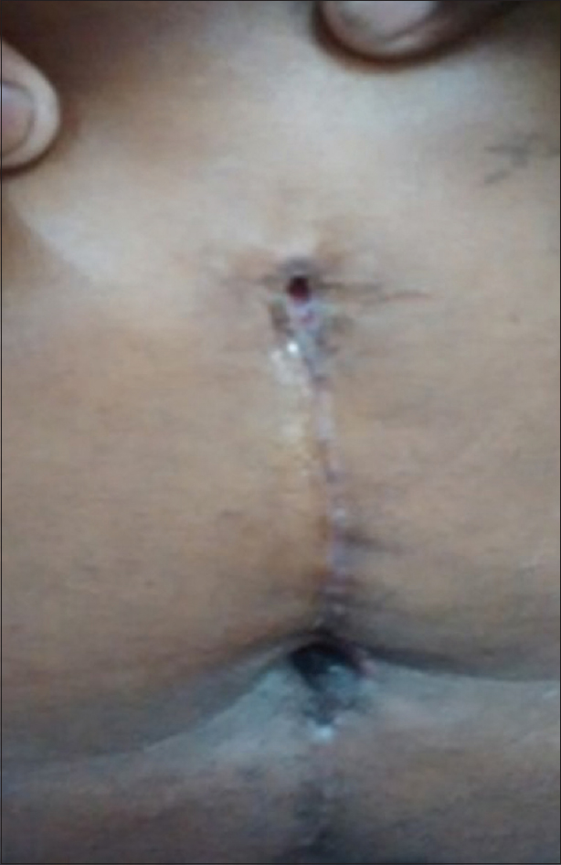
- A case of laparoscopic port site infection following cholecystectomy

- A case of mesh hernioplasty presenting with discharging pus
Modified candle jar technique
Extinction of lighted candle inside an air tight jar leads to consumption of the majority of oxygen inside the jar in a few seconds with residual 1%–2% oxygen and generation of 4%–5% carbon dioxide. The remaining oxygen is then slowly removed by a second step combustion using activated steel wool, resulting in reduction of oxygen inside the jar to the critical level required for the culture of anaerobes.[12]
Agents for combustion
-
Candles
-
Plastic airtight jar: Commercially available, transparent, heat tolerant, gas-impermeable, hard, airtight 1000 ml plastic jar (Tarson, India).
Agents to absorb residual oxygen
Slow oxygen purging
Steel wool (Grade 0–1) 5 gauge à Dip in 50 ml acidified Cu2SO4 solution (10% Cu2SO4 5 ml + Tween 80 10% 5 ml + H2SO4 2 mol/L 3 ml + DW 200 ml) 30–45 s/till steel wool becomes copper coated.
Agents for CO2 generation
A small test tube containing mixture of 0.5 g sodium bicarbonate and 0.5 g magnesium carbonate was kept ready to put inside one 1000 ml test jar, just after placing inoculated plate. On adding water just before closing the lid, chemical reaction will occur leading to carbon dioxide generation.
Anaerobic indicator
Modified methylene blue indicator was used.
Clinical specimens showing acid-fast Bacilli on ZN staining and those obtained from SSIs were inoculated on LJ medium besides MacConkey agar and SBA.
Growths on aerobic culture were identified by conventional morphological and biochemical property study.[13] Identification of anaerobes from culture plates were done by the following methods: The study of colony characteristics, pigment production, fluorescence, Gram-staining, aerotolerance test, biochemical tests including spot indole test, nitrate reduction test, catalase test, sodium polyanethol sulfonate disk test, bile tolerance, lecithinase test, urease production test, sugar fermentation tests, and test for ability to grow against special potency antibiotic discs for anaerobic identifications such as vancomycin (5 µg), colistin, (10 µg) and kanamycin (1 mg).[14]
Growths on LJ media were subjected to ZN staining and on detection of AFB were approached for identification by considering Mpt 64 antigen (for Mycobacterium tuberculosis complex) detection test, period of incubation for growth, pigment production, some conventional biochemical tests such as catalase, peroxidase, aryl sulfatase, niacin accumulation test, and sensitivity to polymixin B. Growth on LJ medium appeared in <1 week. Catalase and aryl sulfatase test were positive, Mpt 64 antigen detection test, peroxidase, and niacin accumulation test were negative and all isolates were resistant to polymixin B. Provisional diagnoses of Mycobacterium abscessus were made. Diagnoses were confirmed by HAIN test which is an RNA-polymerase chain reaction to detect M. tuberculosis complex and few atypical mycobacteria. The test was done in an outside laboratory (Department of Microbiology, Calcutta Medical Research Institute, Kolkata) [Figure 3].

- HAIN test identifying the atypical isolate as Mycobacterium abscessus
Antimicrobial Susceptibility testing for aerobic bacteria was done by Kirby-Bauer disc diffusion method and for anaerobic bacteria by E-test strips incubated in modified candle jar system following CLSI 2014 guidelines.[15] BF production capability of the aerobic organisms was detected by 96 well tissue culture plate method by Stepanovic et al.[16]
Colony BF of atypical Mycobacteria and anaerobes was grown on polycarbonate membrane filter following the methodology of Anderl et al.[17] as follows in brief: 10 μL drops of bacterial suspension grown in BHI broth or Robertson's cooked meat broth (for anaerobes only) was adjusted to 106 CFU/ml and used to seed on black polycarbonate membrane filters (25 mm diameter; pore size 0.22 μm, Millipore, Germany) and was placed on brain–heart infusion agar plates or prereduced Brucella blood agar enriched with hemin and menadione (for anaerobes only). The plates were inverted and incubated at 37°C or in modified candle jar for anaerobes. The membrane-supported BFs were transferred to fresh culture medium in every 24 h. Incubation was done for a total of 14 days for atypical Mycobacteria and 6 days in case of anaerobes. The growths on the membranes were washed with phosphate-buffered saline (PBS, pH 7.2) by agitation at 180 rpm for 1 min to remove nonadherent cells. AFM of the growths was then carried without further sample treatment in the tapping mode in an outside center (Saha Institute of Nuclear Physics, Kolkata, West Bengal, India). We have used the tapping mode as it is suitable for soft materials. In this mode, the stiff cantilever is oscillated at a very close separation to the sample. The height images were analyzed using the WSxM software.[11]
Five lacrimal mucosa samples were included in our study, of which two did not yield any growth on blood agar and MacConkey agar. Histological block was made for them and slides of cross sections were studied by H and E staining,[18] Gram-staining, and fluorescent in situ hybridization technique (FISH).[19] Surface-associated BF on cross sections of lacrimal mucosae was demonstrated using epifluorescence microscopy following FISH.
Fluorescent in situ hybridization protocol for biofilm study (in brief)
The slide was fixed in 4% paraformaldehyde solution for 3 h. Lysozyme enzymatic buffer was added and incubated for 4 h at 45°C. The slide was washed in wash buffer and air dried, followed by incubation in FISH buffer (contains EUB 338 Probe) in a humidity chamber at 55°C for 4 h. Next to it, the slide was washed with wash buffer and air dried and exposed to 10 microliter/ml working concentration of ConA: Alexa fluor488 for 1 h at room temperature followed by Hoechst 33252 stain (2 µg/ml) stain for 1 h at room temperature. All staining were done in dark.[19]
Results
Out of fifty different samples, sixty-three isolates were obtained in pure culture (15 samples showed growth of two organisms, thirty-three samples showed growth of single organism, and two samples of lacrimal mucosae showed no growth which were processed by FISH) as follows: Staphylococcus aureus (25.39%), Escherichia coli (14.28%), Klebsiella pneumonia (14.28%), M. abscessus (12.69%), Citrobacter spp.(9.52%), Bacteroides fragilis (6.3%), Pseudomonas aeruginosa (4.7%), Proteus spp (4.7%), Staphylococcus epidermidis (3.1%), Enterobacter spp. (1.5%), Morganella morganii (1.5%), and Peptostreptococcus spp. (1.5%). Among the isolates, 74% were found to be BF producers in the following frequency: P. aeruginosa 100%, S. epidermidis 100%, B. fragilis 100%, Klebsiella spp. 88.88%, S. aureus 81.25%, M. abscessus 75%, Citrobacter spp. 83.33%, Proteus spp. 66.66%, E. coli spp. 33.33%, Enterobacter spp. 0% [Table 1].

AFM data were collected at 10 × 10 μm scan. Three-dimensional AFM images of colony BF of M. abscessus and Bacteroides fragilis showed cones and domes structure typical of BF polysaccharide [Figures 4 and 5].

- Atomic force microscope surface topography image of colony biofilm of Bacteroides fragilis

- Atomic force microscope surface topography image of colony biofilm of Mycobacterium abscessus
H and E staining of lacrimal mucosae histopathological cross sections showed lakes of polysaccharide [Figure 6]. Gram-staining of the blocks showed colonization by Gram-positive cocci [Figure 7]. FISH done from the lacrimal mucosae blocks showed red green areas of BF colonization [Figure 8].

- H and E image of lacrimal mucosa of a chronic dacryocystitis patient showing polysaccharide lake

- Gram-staining of histological block of lacrimal mucosa showing colonization by Gram-positive cocci

- Fluorescent in situ hybridization of lacrimal sac showing red green areas of biofilm colonization
Discussion
It is a very common clinical experience that in spite of treatment with recommended antibiotics, some of the chronic infections such as anal fistula, chronic dacryocystitis, mesh hernioplasty infections, SSIs, laparoscopic port site infections, osteomyelitis, and chronic nonhealing wounds are known to recur. This is a common feature of BF due to high resistance persisters within exopolysaccharide substances. Hence, we have reviewed such clinical conditions in the light of BF by in vitro studies. In the present study, many of the isolates were found to be BF producers by microtiter tissue culture plate method, in vitro grown colony BF study by AFM, and in vivo by H and E, FISH, and Gram-staining.
From anal fistulae cases, five anaerobic isolates were obtained by an innovative modified candle jar technique and their BF status were demonstrated for the first time by AFM after colony BF formation applying modified candle jar method. Four among the five isolates were BF producers. Strong BF producer S. aureus was isolated in some cases. Viewing anal fistula in the light of BF will help in effective management.
In mesh hernioplasty cases, S. aureus, S. epidermidis, and M. abscessus were isolated. Most isolates were strong BF producers. Clinical history of repeated antibiotic treatment was given by patients not resulting in cure. We advocated removal of mesh under antimicrobial coverage and it was found that cure occurred only after removal of the mesh. Reslinski et al. reports a case of mesh hernioplasty done for an incisional hernia (postlaparotomy scar) patient wherein the presence of a BF was confirmed.[20]
Laparoscopic port site infections showed growth of M. abscessus and S. aureus. All three S. aureus isolates and three out of five M. abscessus isolates were BF producers. For the first time, we detected BF production by atypical mycobacteria by a novel approach of growing in vitro colony BF on polycarbonate membrane followed by detection of BF status by visualization of characteristic surface topography under atomic force microscope. Atypical mycobacteria are well known to form BF as survival strategy as found in different studies.[21]
From chronic osteomyelitis cases S. aureus, M. morganii, and E. coli were isolated, and all of them were BF producers. M. morganii has been a rare inclusion in the list, both as a pathogen for chronic osteomyelitis and a BF producer, as found in this study.[2223] Chronic osteomyelitis cases are known to harbor BF. A study group from the United States have reported BF colonization in osteomyelitis of jaw demonstrated by SEM and histopathology.[24] The main principles of osteomyelitis treatment are debridement of the necrotic tissues in a radical manner, filling up of the dead space, and effective long-term antibiotic therapy which is ideal for BF infection. Frequent investigations and change of antimicrobial therapy should be discouraged.
Many cases of chronic nonhealing ulcer with even no history of immunocompromised state and history of treatment with culture sensitive antibiotics did not result in complete cure. Specimens from these cases showed growth of BF producer isolates of S. aureus and P. aeruginosa. The wounds showed improvement after surgical debridement under prolonged antibiotic treatment which is ideal for BF wound management. In a study by James et al., 60% chronic wounds were found to be BF positive in comparison to 6% acute wounds.[3]
Five chronic dacryocystitis cases were studied. Three of them showed growth of S. aureus and S. epidermidis which were BF producer (by microtiter plate method). No organism was grown in vitro in two cases but showed lakes of polysaccharide on H and E staining. BF persisters may often not grow in cultures. However, fluorescent microscopy was done after staining the histological cross section with Alexa (green), hoechst 33252 (blue tissue stain), and eubacterial stain EUB 338 probe (red) for FISH. Red indicated eubacteria inside the BF matrix which was indicated by green alexa under fluorescent microscope. Gram staining of the histological sections showed colonization by Gram-positive cocci. In a similar study, Oates et al. demonstrated that putative BF production by colonizing microorganisms in cases of chronic diabetic foot ulcers by FISH.[19] In a study reported by Kosarsoy et al., BF was detected by SEM in 12 out of 14 (85.7%) specimens from lacrimal mucosa of cases of chronic dacryocystitis.[25] Culture-directed optimal antibiotic treatment during acute stages may prevent BF colonization. Once colonization has occurred, lacrimal syringing, and dacryocystorhinostomy/dacryocystectomy (mechanical removal of focus) remain the main modality of treatment.
SSIs (such as postopen cholecystectomy, postlaparotomy, and postmastectomy) were often polymicrobial, and the isolates were BF producers. The polymicrobial nature and potential involvement of BFs in SSIs may be analogous to periodontal disease, where a diverse community of microorganisms acting in consort over time results in a chronic infection.[26] Surgical debridement and removal of leftover suture materials (in two cases only) under antibiotic coverage showed improvement in the cases studied followed by complete cure. In a study report, Kanthju et al. showed the presence of Bacilli and cocci within BFs on explanted sutures from a case of chronic SSI by confocal microscopy.[27]
As sessile form is not amenable to therapeutic dose of most sensitive drugs and often require mechanical/chemical curing, rational policy for management of such treatment refractory cases should be planned with destabilizing the colonized surface under cover of antibiotic. However, it is preferred to prevent BF like colonization by early intervention while infective agents are still in planktonic form.
Summary and Conclusion
About 74% of the isolates from chronic discharging sinuses were found to be BF producers. Therefore, chronic discharging sinuses should be viewed in the light of BF, besides other risk factors for chronic infections as the management algorithm would change according to the presence or absence of BF. Antimicrobials tested to be active in vitro, may not be so in vivo, in the face of BF colonization. Mechanical removal under antimicrobial coverage is still the most reliable treatment option for BF infections.
Financial support and sponsorship
Nil.
Conflicts of interest
There are no conflicts of interest.
Acknowledgments
Prof. Sangam Banerjee, Saha Institute of Nuclear Physics, Kolkata - For technical help in AFM Dr. Subhrangshu Mondal, Consultant Microbiologist, CMRI, Kolkata- for technical help in HAIN test Doctors and Staff of Department of Surgery, IPGMER, Kolkata.
References
- Bacterial biofilms: An emerging link to disease pathogenesis. Annu Rev Microbiol. 2003;57:677-701.
- [Google Scholar]
- Effect of surface roughness and materials composition. J Biomater Nanobiotechnol. 2012;3:541-6.
- [Google Scholar]
- Bacterial biofilms: A common cause of persistent infections. Science. 1999;284:1318-22.
- [Google Scholar]
- The application of biofilm science to the study and control of chronic bacterial infections. J Clin Invest. 2003;112:1466-77.
- [Google Scholar]
- Prevention and control of bacterial infections associated with medical devices. Am Soc Artif Intern Organs J. 1992;38:174.
- [Google Scholar]
- A scanning and transmission electron microscopic study of an infected endocardial pacemaker lead. Circulation. 1982;66:1339-41.
- [Google Scholar]
- Anaerobic culture on growth efficient bi-layered culture plate in a modified candle jar using a rapid and slow combustion system. Indian J Med Microbiol. 2013;31:173-6.
- [Google Scholar]
- Mackie and McCartney Practical Medical Microbiology 1988
- Introduction. In: Isenberg HD, ed. Clinical Microbiology Procedures Handbook. Vol 1. Washington, DC: American Society for Microbiology; 2007. p. :4611-46131.
- [Google Scholar]
- Clinical and Laboratory Standards Institute. Performance Standards for Antimicrobial Susceptibility Testing; Twenty-Third Informational Supplement (M100-S23); CLS Document. Pennsylvania, Wayne, PA, USA: Clinical and Laboratory Standards Institute; 2014.
- A modified microtiter-plate test for quantification of staphylococcal biofilm formation. J Microbiol Methods. 2000;40:175-9.
- [Google Scholar]
- Role of antibiotic penetration limitation in Klebsiella pneumoniae biofilm resistance to ampicillin and ciprofloxacin. Antimicrob Agents Chemother. 2000;44:1818-24.
- [Google Scholar]
- Biofilm detection with hematoxylin-eosin staining. Arch Otolaryngol Head Neck Surg. 2010;136:453-6.
- [Google Scholar]
- The visualization of biofilms in chronic diabetic foot wounds using routine diagnostic microscopy methods. J Diabetes Res 2014 2014:153586.
- [Google Scholar]
- In vivo biofilm on the surface of a surgical mesh implant. Pol J Microbiol. 2009;58:367-9.
- [Google Scholar]
- Nontuberculous mycobacteria pathogenesis and biofilm assembly. Int J Mycobacteriol. 2015;4:36-43.
- [Google Scholar]
- Morganella morganii osteomyelitis complicated by secondary septic knee arthritis: A case report. Acta Orthop Traumatol Turc. 2012;46:464-7.
- [Google Scholar]
- Microbial biofilms in osteomyelitis of the jaw and osteonecrosis of the jaw secondary to bisphosphonate therapy. J Am Dent Assoc. 2009;140:1259-65.
- [Google Scholar]
- Presence of biofilms in the lacrimal sac mucosa. Turk Klin J Med Sci. 2013;33:1421-5.
- [Google Scholar]
- Pathogenic synergy: Mixed infections in the oral cavity. Antonie Van Leeuwenhoek. 1984;50:789-98.
- [Google Scholar]
- Chronic surgical site infection due to suture-associated polymicrobial biofilm. Surg Infect (Larchmt). 2009;10:457-61.
- [Google Scholar]


