
Translate this page into:
Case of Lingual Cysticercosis and Review of Literature
Address for correspondence: Dr. Arvind Kinger, E-mail: akinger_73@rediffmail.com
This is an open-access article distributed under the terms of the Creative Commons Attribution-Noncommercial-Share Alike 3.0 Unported, which permits unrestricted use, distribution, and reproduction in any medium, provided the original work is properly cited.
This article was originally published by Medknow Publications & Media Pvt Ltd and was migrated to Scientific Scholar after the change of Publisher.
Abstract
A 30-year-old female presented with a painless solitary swelling at right lateral border of tongue of 2-month duration. Fine-needle aspiration cytology was nonconclusive. Excision biopsy was done. Histopathology revealed cysticercosis cellulosae and parasite visualized in the slide with tongue muscles. Lingual cysticercosis is rare and therefore its literature is reviewed and discussed.
Keywords
Cysticercosis cellulosae
lingual
rare disease

INTRODUCTION
The disease is named after the larval stage of Taenia Solium, cysticercosis cellulosae. The common sites of cysticercosis are skeletal muscles, subcutaneous tissues, and brain. The involvement of the central nervous system causes severe symptoms and can be potentially fatal.[1–3] Although oral involvement is common in swine, it is rare in humans.[45] Literature review has mentioned that only 34 cases of lingual cysticercosis are reported till now.[6] Lingual cysticercosis is rare and not suspected for an isolated swelling over the tongue and therefore produces a diagnostic dilemma for the clinicians. This case therefore is one more addition to this rare group. Excision biopsy was done in this case and the sample was sent for histopathology examination, which confirmed the diagnosis of lingual cysticercosis. We here present a case of isolated lingual cysticercosis without involvement of any other site.
CASE REPORT
A 30-year-old female, presented with painless solitary swelling near right lateral border of tongue of two months duration [Figure 1]. The swelling was progressive in nature, causing discomfort while eating. On examination there was a 3 × 2 × 1 cm well defined pink, smooth, nontender swelling at the junction of anterior one-third and posterior two-third of tongue. General and systemic examinations were within normal limits. Fine-needle aspiration cytology was nonconclusive. Written consent was taken and surgical excision was done under local anesthesia [Figure 2]. It was an encapsulated cystic mass containing yellowish pale fluid [Figure 3]. The postoperative period was uneventful. The histopathological examination revealed cysticercosis cellulosae in tongue musculature [Figure 4]. The diagnosis of lingual cysticercosis was thus established and the patient was advised other investigations to rule out the presence of disease in other parts of the body. CT-scan brain and chest X-Ray were done which confirmed the absence of disease in the respective sites.
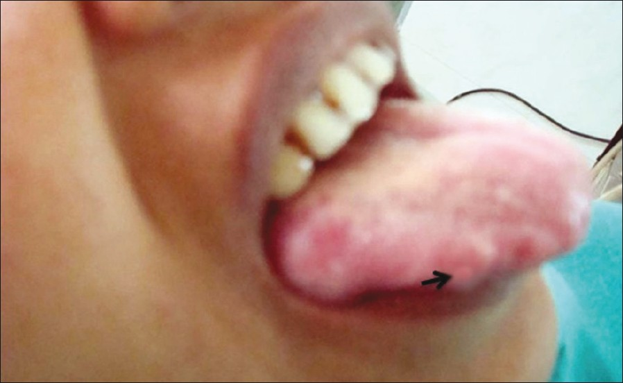
- Swelling right lateral border of tongue (arrow)
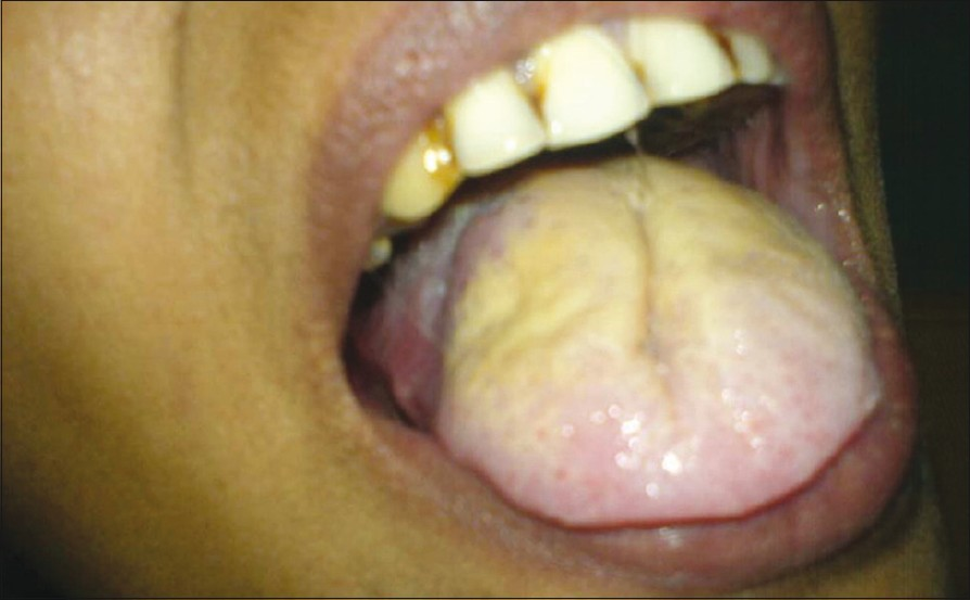
- Post treatment picture
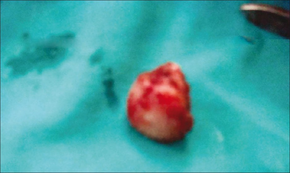
- Excised cyst
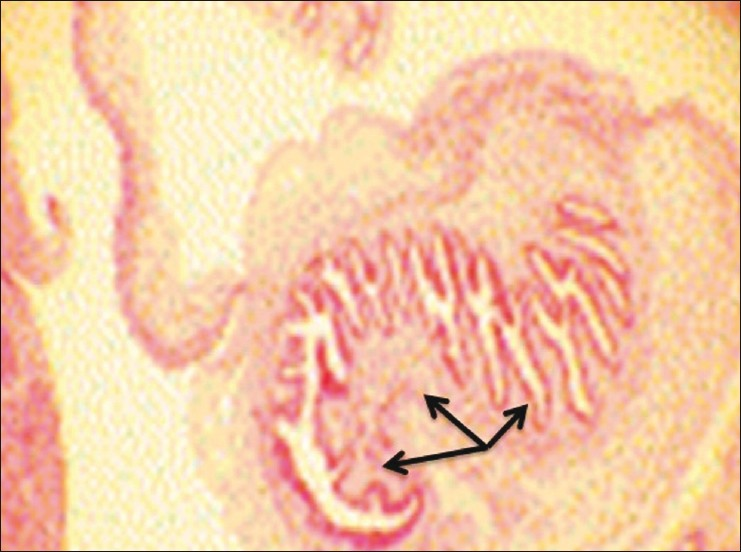
- Cystic cavity containing cysticercus cellulosae larvae (H and E, 10×)
The patient was given Albendazole 200 mg TDS for 30 days. One-year follow-up showed no signs of recurrence.
DISCUSSION
Cysticercosis most frequently infects skeletal muscles, subcutaneous tissue, brain, and ocular tissue.[1–3] The life cycle of Taenia solium involves human as a definite host and pig as an intermediate host. The adult worm lives in small intestine of man measuring 2-3 m in length with 800--900 proglottids. Each gravid segment contains about 50,000 eggs. The worm sheds gravid segments with eggs in the stool infecting pig and man. On reaching the alimentary tract of pig, the eggs rupture, liberating the oncospheres which penetrate the gut wall and reach the systemic circulation. They dislodge into different organs and muscle developing into larvae. Human beings are infected by eating raw contaminated pork containing cysticerci, which develops into adult tapeworm in the jejunum. Autoinfection may also occur by contaminated fingers or direct contact with another human carrier.[57] The worm sheds gravid segments laden with eggs in stool, reinfecting pigs and thus completing the cycle. These are partially digested in stomach, evolving to onchospheres and subsequently penetrate small intestine mucosa to disseminate throughout the body via arteriovenous and lymphatic channels. The clinical manifestations depend on location and number of cysticercosis at particular site. Intestinal infection by Taenia solium could be asymptomatic or may present with epigastric pain, nausea, and loose motions. In the case of neurocysticercosis, a patient presents with seizures and the other symptoms are related to the location and pressure caused by cysticerci in particular the region of the brain. Oral and subcutaneous forms are asymptomatic but are detected early because of their superficial location.[8] Diagnosis is confirmed by the presence of cysticerci on biopsy. Roentgenograms of skull and soft tissues may reveal calcified cysticerci. Eosinophilia may occur in invasive stage.
The treatment depends on symptoms and accessibility of lesion. Neurocysticercosis and multiple cysts are treated with drugs like Praziquantel and Albendazole.[9] Treatment of choice in solitary accessible lesion is surgical excision.[910]
Source of Support: Nil.
Conflict of Interest: None declared.
REFERENCES
- Oral cysticercosis: Case report. Oral Surg Oral Med Oral Path Oral Radol Endod. 2007;104:56-8.
- [Google Scholar]
- Oral cysticercosis: A collaborative study of 16 cases. Oral Surg Oral Med Oral Path Oral Radol Endod. 2007;103:528-33.
- [Google Scholar]
- Oral cysticercosis. Oral Surg Oral Med Oral Pathol Oral Radiol Endod. 1995;79:572-7.
- [Google Scholar]
- Helminthic diseases. In: Oral diseases in the tropics. Oxford: Oxford Medical Publications; 1993. p. :126-9.
- [Google Scholar]
- Oral cysticercosis: Review of literature and report of two cases. Int J Oral Surg. 1981;10:371-5.
- [Google Scholar]
- Cysticercosis of the oral cavity: Report of five cases and a review of literature. Int J Pediatr Dent. 1998;8:273-8.
- [Google Scholar]
- Cysticercosis of the oral cavity- A clinicopathological study of ten and a half years. J Indian Dent Asso. 1986;58:257-9.
- [Google Scholar]


