
Translate this page into:
Comparison of Microwave Versus Conventional Decalcification of Teeth Using Three Different Decalcifying Solutions
Address for correspondence: Dr. Archana Srinivasyaiah, E-mail: archana.srinivas20@gmail.com
This is an open access article distributed under the terms of the Creative Commons Attribution NonCommercial ShareAlike 3.0 License, which allows others to remix, tweak, and build upon the work non commercially, as long as the author is credited and the new creations are licensed under the identical terms.
This article was originally published by Medknow Publications & Media Pvt Ltd and was migrated to Scientific Scholar after the change of Publisher.
Abstract
Background:
In routine histopathology, decalcification of teeth is an essential and important step during tissue processing. The present study was attempted to decalcify teeth using microwave method and to compare it with conventional decalcification method.
Aim:
To compare microwave decalcification with conventional decalcification method with respect to the speed of decalcification, preservation of tissue structure, and efficacy of staining.
Materials and Methods:
A total of 72 single-rooted premolars were used for both conventional and microwave decalcification methods. Three different types of decalcifying agents at 5% and 7% were used. Decalcifying agents included nitric acid, formic acid, and trichloroacetic acid, each at 5% and 7%. About 6 teeth were included in each category and were decalcified by both conventional and microwave method. After decalcification, all the teeth were examined macroscopically and microscopically. Cramer's V-test was used to determine the statistical significance.
Results:
The results have shown that microwave method using 5% and 7% nitric acid were the fastest. Structural details and good staining characteristics were better in teeth decalcified by 5% nitric acid and 5% trichloroacetic acid by both the methods.
Conclusions:
5% nitric acid by microwave method proved to be the best decalcifying agent as it was fast and gave good structural details and staining characteristics.
Keywords
Conventional
decalcification
formic acid
microwave
nitric acid
premolars
teeth
trichloroacetic acid

INTRODUCTION
Hard tissue preservation close to the viable state is indispensable for the understanding of structural and sub-structural details and functions. The cutting of thin sections by conventionally used methods is impossible in cases of tissues such as bone, teeth, odontomas, and calcified lesions. For such tissues, removal of calcium is done by a process known as “decalcification” which in turn makes the tissue soft enough to be cut by the microtome.[1]
Hard tissue decalcification is one of the most sensitive procedures in the histopathology laboratory. It is of importance, especially in the field of oral pathology, as teeth decalcification is a routinely required procedure.[2] Decalcification is generally carried out by chemical agents, either with inorganic acids to form soluble calcium salts or chelating agents that bind to calcium ions.[3]
In the conventional method of decalcification, hard tissue is placed in a decalcifying agent at room temperature with changes of the solution at regular intervals till the decalcification end point is reached. Microwave method of decalcification is a novel method compared with the conventional method. By the use of this method, hard tissues are placed in the decalcifying agent in a microwave oven for intermittent periods with regular changes of the solution till the end point is reached. Microwave irradiation of the hard tissue specimens has shown to speed up the decalcification process significantly from days to hours.[4]
Mayers was the first person who recognized the application of microwave energy in histotechnology in 1970. Microwaves are nonionizing radiation which produces alternating electromagnetic fields which in turn result in the rotation of dipolar molecules such as water and the polar side chains of the proteins through 180°C at the rate of 2.45 billion cycles per second. This induces molecular kinetics which results in the generation of energy flux which continues until the radiation ceases.[5] Research has shown that bone decalcification by microwave method is accelerated about 10 times when compared with the conventional method, at ambient temperature.[6]
The present study was aimed to study decalcification of teeth by conventional method and microwave method using three different decalcifying agents each at two different percentages, and to find the best decalcifying solution and percentage to be used with respect to the speed of decalcification, preservation of tissue structure, and staining efficacy in both the methods.
MATERIALS AND METHODS
A total of 72 extracted single-rooted premolar teeth were used in the study. They were divided equally into two groups based on the method by which the teeth were decalcified; Group 1: Conventional decalcification method and Group 2: Microwave decalcification method. Three different acids at two different concentrations were used as the decalcifying agents, i.e. nitric acid 5% and 7%, formic acid 5% and 7%, and trichloroacetic acid 5% and 7%. About 6 teeth were decalcified in each category. Kitchen microwave oven of LG Company with 800 W, model 2049 W, which was placed in stainless steel chamber fitted with vent for fumes released by reagents to escape outside the laboratory, was used for microwave decalcification [Figure 1]. The results were recorded and statistically analyzed.
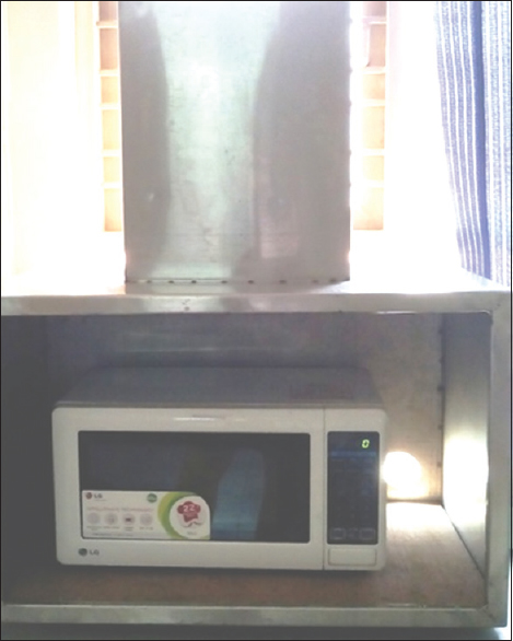
- Microwave oven placed in a stainless steel enclosure fitted with vent opening to the exterior environment
Decalcification methodology
All the samples were properly labeled, and each sample was suspended in a beaker containing the decalcifying solution, with the help of thread for conventional and microwave decalcification. The exact time at the start of decalcification was noted. The end point of decalcification was determined by chemical method using calcium oxalate method (Clayden 1952 - Chemical method). During public holidays and weekends, the tooth samples under the process of decalcification were maintained as “status quo.” In microwave method, the beaker containing the decalcification solution and the tooth specimen were placed in a microwave oven and irradiated for 2 min. Eight cycles of such irradiation per day, with 1 h interval was carried out for all the three solutions. The temperature of all the three solutions was maintained at around 41–43°C. Macroscopically, yellow discoloration of specimen, tearing, and shrinkage were assessed. Specimens were then processed; sectioned and stained with hematoxylin and eosin. Microscopically, sections were assessed for the quality of staining and overall structural detail. The macroscopic parameters were recorded as present or absent, and the microscopic parameters were graded as good, fair, and poor.
The data obtained from the above observations were entered and analyzed using SPSS version 21 software, IBM Nitric, Formic & Trichloroacetic acid) are by Nice Chemicals Pvt. Ltd., Kochi, Kerala. Descriptive statistics was used for all parameters, and Cramer's V-test was applied for significance to the collected data.
RESULTS
Out of the two methods which were used, microwave method of decalcification was faster than the conventional method. Using microwave method, 5% and 7% nitric acid took 2 days to decalcify tooth structure which was followed by 7% and 5% trichloroacetic acid which took 13 days and 18 days, respectively, and the maximum time of decalcification was taken by 7% and 5% formic acid which were 20 and 21 days, respectively. By the conventional method, 7% nitric acid took the least time of 12 days and 5% formic acid took the maximum time of 42 days. Hence, the process of decalcification was made faster by microwave method and was found to be statistically significant (P < 0.05) [Table 1].

Discoloration of teeth
Discoloration of teeth was seen only when 5% and 7% nitric acid was used as decalcifying agent with both the methods [Figure 2] whereas the other two acids did not produce any discoloration. Hence, nitric acid used in either of the percentages irrespective of the methods used will produce tooth discoloration in comparison with other decalcifying agents, which was statistically significant [Table 2].
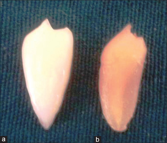
- Discoloration: Yellow color change in the tooth after decalcification (b) with nitric acid in comparison with the tooth before decalcification (a)

Tearing and shrinkage of teeth
Formic acid and trichloroacetic acid showed tearing and shrinkage in either of the percentages by both conventional and microwave method, which was statistically significant [Table 3 and Figure 3]. 7% nitric acid produced tearing and shrinkage, which was seen in minimal number of teeth.

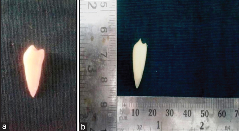
- Comparison of tearing and shrinkage of tooth before (a) and after (b) decalcification with formic acid
Overall structural details
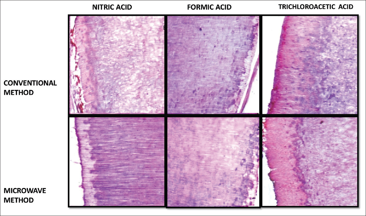
Among all the decalcified teeth, majority of the teeth showed good structural details by microwave method. In conventional method, the structural details ranged from good, fair, and poor [Figures 4 and 5]. Among the decalcifying agents, 5% nitric acid and 5% and 7% trichloroacetic showed good details by microwave method and 5% nitric acid and 7% trichloroacetic acid showed good details by conventional method, but these findings were not statistically significant [Table 4].
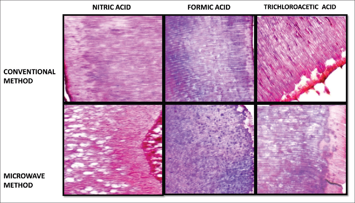
- H and E, section revealing staining characteristics and structural details for 7% nitric acid, formic acid, and trichloroacetic acid using conventional and microwave method (×100)
- H and E, section revealing staining characteristics and structural details for 5% nitric acid, formic acid, and trichloroacetic acid using conventional and microwave method (×100)

Overall staining quality
In microwave method, 5% nitric acid showed good staining characteristics, 5% and 7% formic acid and 7% trichloroacetic acid showed fair staining characteristics. In conventional method, 5% trichloroacetic acid has shown good overall staining of the tissue and 5% and 7% formic acid and 7% trichloroacetic acid has shown fair staining characteristics. Out of all the decalcified teeth, microwave method of decalcification showed significantly better staining characteristics than conventional method [Table 5 and Figures 4, 5].

DISCUSSION
The decalcification of teeth is done to study the tooth structure, pulp calcifications, and also to evaluate the biological response of dental pulp to restorative materials.[7] Research has been done on the introduction of new decalcifying agents as well as modification of known decalcifying agents[3] to meet the criteria of a good decalcifying agent which should ensure calcium removal completely at a reasonable speed with minimal damage to cells and tissues.[8]
Tooth decalcification is a time-consuming process. It takes days to weeks, and tissue structure preservation depends on the quality of the demineralization process. A new method using microwave oven was seen to increase the speed of decalcification process.[9] Depending on the urgency, decalcifying agent and method are decided.[10]
A study done on decalcification by Pitol et al. using microwave method has shown that there was 30-fold increase in decalcification speed with microwave method as compared with the conventional method. Another study has shown a reduction of decalcification time from 45 days to 2 days when done by microwave method.[9] Others opine that the bone decalcification is accelerated by about 10 times using microwave method.[6] The results of the present study were consistent with the previous studies, concluding that the microwave-assisted method is faster than the conventional method.
According to a study done by Moore et al.,[11] Lynch[1] and Culling et al.,[7] yellow staining of the tissue results when nitric acid is used as a decalcifying agent. In our study, we also observed that nitric acid produced yellowish discoloration of teeth when used as a decalcifying agent irrespective of the method used. The other decalcifying agents did not produce any discoloration of teeth. Formic acid and trichloroacetic acid produced tearing and shrinkage of teeth in both the microwave and conventional methods. This fact is not well documented in the scientific literature, but as per our study, we found that these decalcifying agents produced tearing and shrinkage of teeth irrespective of the method used.
Ethylene di-amine tetra acetic acid (EDTA) and formic acid, irrespective of the method used have shown good overall structural details.[12] Our finding varied from the other study in that we did not consider EDTA in the present study. Second in our study, formic acid decalcification did not give good results by microwave method and 5% nitric acid and 5% trichloroacetic acid gave the best results by both the methods.
Regarding staining characteristics, our study showed that nitric acid and trichloroacetic acid when used by either of the methods produced good staining characteristics which were consistent with a study done by Sangeetha et al.[12]
CONCLUSION
Decalcification of hard tissue specimen is a very technique sensitive method and plays an important role in oral pathology, as decalcification of bone or teeth is done on regular day-to-day basis for histopathology reporting. To obtain paraffin or celloidin sections of hard tissue, calcium must be removed to study hard tissue. Conventional decalcification method is practiced regularly which gives good structural details, but it takes a long time to decalcify leading to delay in histopathology reporting. With our study, we can conclude that microwave-assisted method of decalcification is faster than conventional method, irrespective of the decalcifying agents used. Although nitric acid produces discoloration of teeth, it does not produce any tissue tearing or shrinkage and gives better structural details and staining characteristics than formic acid or trichloroacetic acid by microwave method. These results are comparable to the conventional decalcification method. Therefore, we propose the use of nitric acid (5%) as decalcifying agent by microwave method routinely in histopathology laboratories.
Financial support and sponsorship
Nil.
Conflicts of interest
There are no conflicts of interest.
Acknowledgment
I would like to thank Dr. Bhuvan Nagpal, my batchmate for his support during the course of my study.
REFERENCES
- Lynch's Medical Laboratory Technology. (4th ed). London: W B Saunders; 1983. p. :937-44.
- [Google Scholar]
- Orban's Oral Histology and Embryology. (10th ed). New Delhi: CBS Publishers and Distributors; 1990. p. :349-54.
- [Google Scholar]
- Theory and Practice of Histological Methods. (6th ed). Edinburgh: Churchill Livingstone; 2008. p. :338-60.
- [Google Scholar]
- Microwave oven for improved tissue fixation and decalcification. Pathologica. 1991;83:307-10.
- [Google Scholar]
- Microwave histoprocessing versus conventional histoprocessing. Indian J Pathol Microbiol. 2008;51:12-6.
- [Google Scholar]
- Evaluation of efficacy of various chemicals for decalcification of dental hard tissues – An in-vitro study. J Orofac Sci. 2010;1:1-10.
- [Google Scholar]
- Microwave-induced fast decalcification of rat bone for electron microscopic analysis: An ultrastructural and cytochemical study. Braz Dent J. 2007;18:153-7.
- [Google Scholar]
- Longitudinal and quantitative evaluation of dentin demineralization when subjected to EDTA, EDTAC, and citric acid: A co-site digital optical microscopy study. Oral Surg Oral Med Oral Pathol Oral Radiol Endod. 2008;105:391-7.
- [Google Scholar]
- Bone. In: Woods AE, Ellis RC, eds. Laboratory Histopathology. New York: Churchill Livingstone; 1994. p. :7-10.
- [Google Scholar]
- Comparison of routine decalcification methods with microwave decalcification of bone and teeth. J Oral Maxillofac Pathol. 2013;17:386-91.
- [Google Scholar]


