Translate this page into:
Disseminated Histoplasmosis: A Fatal Opportunistic Infection in Disguise
Address for correspondence: Dr. Ruchita Tyagi, E-mail: ruchitatyagi@gmail.com
This is an open access article distributed under the terms of the Creative Commons Attribution NonCommercial ShareAlike 3.0 License, which allows others to remix, tweak, and build upon the work non commercially, as long as the author is credited and the new creations are licensed under the identical terms.
This article was originally published by Medknow Publications & Media Pvt Ltd and was migrated to Scientific Scholar after the change of Publisher.
Abstract
Histoplasma capsulatum is no longer confined to certain geographic areas and should always be considered in the differential diagnosis of lymphadenopathy and organomegaly in HIV-positive patients. We present an unusual case of a 20-year-old immunocompromised male of African origin presenting with fever, jaundice, hepatosplenomegaly, and retroperitoneal and cervical lymphadenopathy. Fine needle aspiration (FNA) smears from the cervical lymph node revealed numerous yeast forms of histoplasma in macrophages. The patient succumbed to the fulminant infection. Postmortem liver biopsy also revealed infiltration by histoplasma, confirming the diagnosis of disseminated histoplasmosis. This case highlights the variable nature of the clinical presentation of disseminated histoplasmosis which can mimic tuberculosis, leishmaniasis, or lymphoma. FNA cytology is a rapid, cost-effective, and reliable diagnostic tool for early detection and prompt management of histoplasmosis.
Keywords
Disseminated
histoplasmosis
immunocompromised
INTRODUCTION
Histoplasmosis, is a fungal infection caused by histoplasma capsulatum, a dimorphic fungus with two distinct varieties affecting the humans: Histoplasma capsulatum var capsulatum and histoplasma capsulatum var. duboisii. The former variety is commonly found in eastern United States (Ohio, Mississippi and St. Lawrence River valleys) and Latin America, with sporadic cases also being reported worldwide. The latter variety, presenting as cutaneous infection, is limited to Sub-Saharan Africa.[1] Immunosuppressed individuals like AIDS patients are at particularly high-risk of developing disseminated histoplasmosis due to ineffective cell-mediated immunity.[2] In fact, histoplasmosis is one of the AIDS-defining illnesses.[3] In India, this opportunistic infection is still an uncommon entity with isolated case reports of disseminated histoplasmosis affecting immunocompromised patients.[45678] The clinical picture may be mistaken for tuberculosis or even lymphoma which are more commonly encountered. Therefore, a high level of clinical suspicion is required to diagnose this uncommon opportunistic infection in nonendemic areas. We present a case of disseminated histoplasmosis in a HIV - positive patient, diagnosed on fine needle aspiration cytology (FNAC) from a cervical lymph node.
CASE REPORT
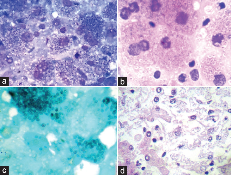
A 20-year-old African male presented with fever and abdominal pain for 15 days and jaundice for 3 days. On examination, he was found to have bilateral cervical lymphadenopathy. On ultrasonographic examination of the abdomen, hepatosplenomegaly, ascites, and retroperitoneal lymphadenopathy were noted. A differential diagnosis of tuberculosis, leishmaniasis, and lymphoproliferative disorder. Examination of the peripheral blood revealed thrombocytopenia (25,000/μl) and leukopenia (3800/μl). FNA was done from the cervical lymph nodes, and smears showed sheets of foamy macrophages containing numerous yeast forms of histoplasma, highlighted on Gomori methenamine silver (GMS) and periodic acid-Schiff (PAS) stains [Figure 1a–c]. During the course of investigations, his serum sample also tested reactive for HIV. As the patient succumbed to severe infection within 1 day of hospital admission, further tests such as culture and CD4 counts could not be done. Postmortem Tru-Cut liver biopsy was performed, which revealed dense infiltration by histoplasma within macrophages [Figure 1d] confirming the diagnosis of disseminated histoplasmosis.

- (a) Fine needle aspiration smear showing yeast forms of histoplasma lying extracellularly and intracellularly within macrophages on Giemsa, ×1000, (b) PAS, ×1000, (c) GMS, ×1000, (d) liver biopsy showing dense infiltration by yeast forms on H and E, ×400
DISCUSSION
Histoplasmosis spreads by inhalation of mycelial fragments and microconidia which are deposited in alveoli.[7] At 37°C in vitro and in tissues, the spores of this dimorphic fungus germinate into yeast forms which are ingested by pulmonary macrophages. The yeasts become parasitic, multiply within these cells, and travel to hilar and mediastinal lymph nodes, where they enter the blood circulation and disseminate to various organs. Macrophages throughout the reticuloendothelial system ingest and sequester the organism. In immunocompetent hosts, T lymphocytes activate macrophages to kill this fungus. However, some organs may still harbor foci of viable histoplasma capsulatum, which is controlled but not completely killed by the immune system. In this way, infection may be reactivated whenever the host faces immunosuppression which may be due to corticosteroids, immunosuppressive drugs, HIV, or any other cause. The deficient cell-mediated immunity is unable to control the organism, resulting in symptomatic infection.[2]
In the absence of serological tests, FNAC serves as a reliable investigation for quick and accurate diagnosis of this infection. The histoplasma yeast forms are identified easily on FNAC smears because of their characteristic appearance. On morphology, the oval, narrow-based budding yeast forms are 2–4 μm in size, which may be present extracellularly as well as intracellularly within macrophages. These may be confused with LD bodies, but the presence of kinetoplasts in leishmania helps to differentiate between these two infections. On routine hematoxylin and eosin stain, it is difficult to detect the tiny yeast forms unless they are present in large numbers. Special stains such as GMS and PAS highlight the yeast forms on cytological smears as well as on the tissue sections, thus helping to arrive at the correct diagnosis.
Histoplasmosis is not a common infection in the Indian scenario. The clinical features of fever, jaundice, hepatosplenomegaly, lymphadenopathy, and ascites are also seen in tuberculosis, leishmaniasis, and lymphoma. In a HIV-positive patient from an endemic area, presenting with organomegaly and generalized lymphadenopathy, the possibility of histoplasmosis should be considered.
CONCLUSION
This case highlights that clinical presentation of disseminated histoplasmosis can be deceptive and may mimic tuberculosis or lymphoproliferative disorder. Histoplasmosis has become more widespread than expected. Lymph node FNAC is a simple, minimally invasive, cost-effective, and reliable diagnostic tool to detect histoplasmosis and hasten the initiation of treatment when time is of essence. The identification of organisms in cytological material permits application of early antifungal treatment before culture results are available.
Financial support and sponsorship
Nil.
Conflicts of interest
There are no conflicts of interest.
REFERENCES
- Histoplasma capsulatum: More widespread than previously thought. Am J Trop Med Hyg. 2014;90:982-3.
- [Google Scholar]
- Histoplasmosis: A clinical and laboratory update. Clin Microbiol Rev. 2007;20:115-32.
- [Google Scholar]
- Disseminated histoplasmosis in patients with AIDS in Panama: A review of 104 cases. Clin Infect Dis. 2005;40:1199-202.
- [Google Scholar]
- Acute Disseminated Histoplasmosisin a patient with AIDS. Indian J Med Microbiol. 1996;14:23-4.
- [Google Scholar]
- Disseminated histoplasmosis with conjunctival involvement in an immunocompromised patient. Indian J Sex Transm Dis. 2010;31:35-8.
- [Google Scholar]
- Histoplasmosis in eastern India: The tip of the iceberg? Trans R Soc Trop Med Hyg. 1999;93:540-2.
- [Google Scholar]


