Translate this page into:
Surgical management of chronic hyperplastic candidiasis refractory to systemic antifungal treatment
Address for correspondence: Dr. Neha Shah, EC-11 Saltlake City, Sector-I, Kolkata - 700 064, West Bengal, India. E-mail: docneha10@gmail.com
-
Received: ,
Accepted: ,
This is an open access article distributed under the terms of the Creative Commons Attribution-NonCommercial-ShareAlike 3.0 License, which allows others to remix, tweak, and build upon the work non-commercially, as long as the author is credited and the new creations are licensed under the identical terms.
This article was originally published by Medknow Publications & Media Pvt Ltd and was migrated to Scientific Scholar after the change of Publisher.
Abstract
Chronic hyperplastic candidiasis (CHC), earlier known as candidal leukoplakia, is a variant of oral candidiasis that classically presents as a white patch on the commissures of the oral mucosa and it is mostly caused by Candida albicans. Clinically, the lesions are usually asymptomatic and regress after appropriate antifungal therapy and correction of the underlying cause. If the lesions are untreated, a small portion may develop dysplasia and later progress into carcinoma. The purpose of this article is to report a case of CHC in a 57-year-old male patient with a significant smoking habit, who presented with a thick, nonscrapable, brownish-white coating on the dorsum of the tongue for 9 years. This case is of particular importance and concern because of the high risk for malignant transformation in CHC. The role of biopsy and histopathology is also stressed through this case report in arriving at a definitive diagnosis and treatment planning. Further, this case is interesting because it was refractory to local and systemic antifungal treatment and so, surgery was chosen as an alternative treatment modality considering the side effects of the prolonged use of antifungal drugs.
Keywords
Chronic hyperplastic candidiasis
malignant transformation
oral candidiasis
smoking
tongue
Introduction
Oral candidiasis (OC) is one of the common infections encountered by the clinicians in the oral cavity, mostly caused by Candida albicans, which is a normal commensal of the oral cavity, skin, gastrointestinal tract, and genitourinary tract.[123] Pseudomembranous, erythematous, and chronic hyperplastic are the three types of primary OC, out of which chronic hyperplastic candidiasis (CHC) is the least common.[245] Although Candida is more frequently isolated from women, the highest prevalence of CHC is seen in middle-aged men who are smokers.[6] CHC is of two types: Homogeneous/plaque and nodular/speckled type. Homogeneous type presents as white plaque that cannot be scraped off and is usually asymptomatic. Speckled type presents as multiple white nodules on an erythematous background and may be associated with pain and burning sensation.[37] The most commonly affected site is the buccal commissure followed by buccal mucosa, palate, and tongue.[78] CHC is of particular significance and concern because of its reported association with malignant transformation; hence, biopsy is mandatory for accurate diagnosis.[37910] Various treatment modalities have been suggested for its management ranging from topical and systemic antifungal agents to surgical methods.[7]
Case Report
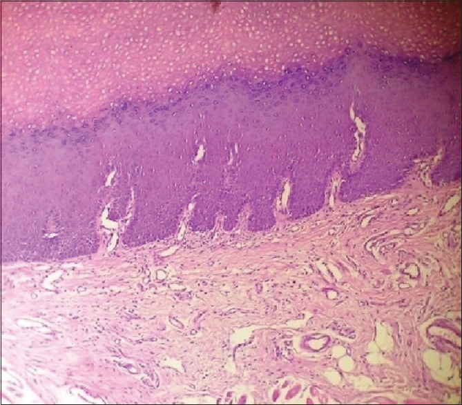
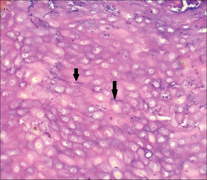
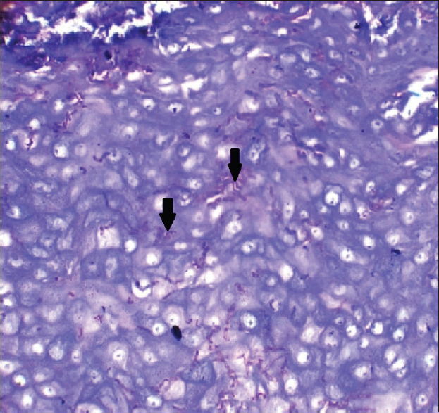
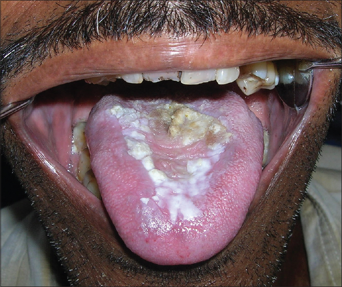
A 57-year-old male patient reported to the Department of Oral and Maxillofacial Pathology of Dr. R. Ahmed Dental College and Hospital, Kolkata, with a chief complaint of a thick coating on the tongue causing discomfort for the past 9 years. His past medical history and family history were noncontributory. He consulted a local physician for his ailment and received several courses of topical and systemic medications including antifungal drugs, without much relief of symptoms. Thereafter, he was referred to our hospital for further management. The patient belonged to poor socioeconomic strata and was a chronic smoker with a history of smoking 1–2 bundles of beedi/day for 15–20 years. Extraoral examination did not reveal any abnormality. Intraoral examination of the patient revealed poor oral hygiene and the presence of thick, brownish-white coating intermixed with erythematous areas, measuring about 4 cm × 2.5 cm on the dorsal surface of the tongue [Figure 1]. The lesion was raised, uneven, granular, and nodular at some places and homogeneous at other areas. The periphery of the lesion was white and merged with the normal mucosa of the dorsum of the tongue. It was nonscrapable, firm, and adherent to the underlying tissue. No similar lesion was present in other areas of the oral cavity including the palate. Based on the history and clinical examination, a provisional diagnosis of CHC was made. Differential diagnosis included oral leukoplakia, proliferative verrucous leukoplakia, oral hairy leukoplakia, hairy tongue, and malignancy. A complete hemogram was done, and all the values were found to be within the normal range. An incisional biopsy was performed and sent for histopathologic examination. Hematoxylin and eosin (H and E)-stained section showed the presence of hyperplastic stratified squamous epithelium with a thick layer of hyperparakeratinization on the surface [Figure 2]. Fungal hyphae were seen on the parakeratin layer [Figure 3]. There was an elongation of the epithelial rete ridges. The underlying connective tissue revealed a dense chronic inflammatory cell infiltrate chiefly comprising lymphocytes. Based on the histopathological examination, a final diagnosis of candidiasis was concluded. Further, periodic acid-Schiff (PAS) staining was done to confirm the presence of candidal hyphae, which were stained magenta red [Figure 4].

- Intraoral photograph showing thick brownish-white coating on the dorsal surface of the tongue with bilateral involvement having uneven, raised, granular areas at some places and homogeneous white patches in the periphery

- Low-power photomicrograph (H and E, ×10) showing hyperplastic epithelium with hyperparakeratosis and elongated rete ridges and chronic inflammation of the underlying connective tissue

- High-power photomicrograph (H and E, ×40) showing candidal hyphae (black arrows) in epithelium

- High-power photomicrograph (PAS, ×40) showing candidal hyphae (black arrows) stained magenta red
Subsequently, the patient was counseled to quit smoking habit and prescribed oral antifungal fluconazole 200 mg once daily along with topical antifungal clotrimazole five times daily for 2 weeks. Following the antifungal therapy, the lesion regressed substantially. However, nonscrapable, adherent, homogeneous white patches remained on the dorsum of the tongue [Figure 5]. Surgery was then done to excise the remaining adherent white patches from the dorsum of the tongue. No lesions were evident in the follow-up period of 6 months [Figure 6].

- Intraoral photograph showing partial resolution of the lesion 2 weeks after antifungal therapy

- Intraoral photograph showing follow-up of the patient after 6 months
Discussion
According to the proposed revised classification of OC by Axéll et al. in 1997, the important forms of primary OC are pseudomembranous, erythematous, and CHC.[25] CHC is the least common form of this opportunistic fungal infection.[24]
Cernea et al. and Jepsen and Winter in 1965 first reported that many oral leukoplakias were associated with Candida infection.[71112] Mehta et al. reported 0.2–4.9% prevalence of oral leukoplakia in India.[13] Interestingly, 7–50% of the leukoplakias have been found to be associated with candidal hyphae.[7] Lehner first used the term “candidal leukoplakia” for leukoplakic lesions associated with chronic candidal infection. However, this term is no longer used; instead, it is designated as CHC.[710]
In the present case, a 57-year-old male patient with a significant smoking habit was diagnosed with CHC, which is in accordance with literature where it has been reported in age group from 31 years to 81 years, though most patients are above 50 years. Males are affected more frequently than females in a ratio of 2:1.[7] In our case, tongue was involved which is an unusual site for occurrence of CHC. The most common being buccal commissure followed by buccal mucosa and palate.[78]
Smoking has been recognized as the main risk factor associated with CHC.[214] Daftary et al. reported that 98% of the patients affected by CHC either smoked or chewed tobacco.[15] Shin et al. have reported a positive and direct association between smoking and candidal colonization.[16] It has been suggested that smoking favors candidal colonization by causing localized epithelial alterations such as increased epithelial keratinization and hydrophobicity. An alternative hypothesis states that smoking depresses the activity of polymorphonuclear leukocytes and reduces gingival exudate, thereby decreasing the number of leukocytes and salivary immunoglobulin A levels which, in turn, are important in inhibiting the candidal colonization in the oral cavity.[17]
Both homogeneous/plaque and nodular/speckled type of CHC can progress to moderate-to-severe dysplasia or even malignancy.[34891014] It has been reported that up to 15% of the CHC lesions may progress to dysplasia.[7] Although the role of Candida in oral carcinogenesis is not well established, in vitro studies have reported the production of carcinogenic nitrosamine, N-nitrosobenzylmethylamine, by Candida from precursors.[610] In 1989, Field et al. postulated that the nitrosamine produced by Candida may directly or along with other carcinogens, activates specific proto-oncogenes responsible for carcinogenesis.[17] In addition, the expression of p53 tumor suppressor gene in cases of CHC has been studied, thereby linking the association of CHC with epithelial dysplasia or malignancy.[7]
Mere presence of Candida in the oral cavity is not indicative of infection, since it is a common inhabitant in this location being present in around 40–60% of the normal individuals.[1] Definitive diagnosis of OC requires the confirmation of tissue invasion by Candida. Hence, biopsy which is considered the gold standard for diagnosis is a must, since the nonscrapable white plaques of CHC are most often confused with leukoplakia clinically.[9] In our case too, biopsy guided in establishing the confirmatory diagnosis and treatment planning.
Histopathological examination of tissue sections stained with H and E reveals epithelial hyperplasia with hyperparakeratosis and candidal hyphae in parakeratin layer invading the deeper layers of epithelium, as was seen in the present case. An inflammatory cell infiltrate consisting of polymorphonuclear leukocytes and lymphocytes is present within the lamina propria and occasionally in the superficial layer of epithelium. The presence of fungal hyphae can be confirmed with PAS stain.[36] Nagai et al. have concluded that epithelial hyperplasia is a protective response of the host against Candida.[18]
Management of CHC involves the elimination of predisposing factors, chiefly smoking, antifungal therapy (both topical and systemic), and surgery in cases nonresponsive to medical treatment.[7] The patient had been complaining of thick coating on the tongue for 9 years and also had received several courses of antifungal drugs by a local physician, without much relief of symptoms. Hence, he was initially prescribed both oral and topical antifungal drugs for 2 weeks only, since the dose and duration of systemic treatment are reduced when a combination of both is used, but when the lesion did not regress completely following the antifungal treatment, surgery was performed to reduce the risk of malignant transformation associated with CHC.
Conclusion
Although OC is a common infection in humans, yet it can pose a diagnostic challenge due to its varied clinical presentations. CHC is the least common form of OC and closely mimics many clinical conditions. Since there is a risk of malignant transformation associated with CHC, histopathological examination is essential to provide conclusive diagnosis and plan treatment.
Financial support and sponsorship
Nil.
Conflicts of interest
There are no conflicts of interest.
References
- Oral candidiasis mimicking an oral squamous cell carcinoma: Report of a case. Gerodontology. 2012;29:70-4.
- [Google Scholar]
- Oral and Maxillofacial Pathology (3rd ed). India: Elsevier Publishers; 2009. p. :217-21.
- A proposal for reclassification of oral candidiasis. Oral Surg Oral Med Oral Pathol Oral Radiol Endod. 1997;84:111-2.
- [Google Scholar]
- Pathogenesis and treatment of oral candidosis. J Oral Microbiol. 2011;3:5771. DOI: 10.3402/jomv.3i0.5771
- [Google Scholar]
- Chronic hyperplastic candidosis/candidiasis (candidal leukoplakia) Crit Rev Oral Biol Med. 2003;14:253-67.
- [Google Scholar]
- Chronic hyperplastic candidiasis: A pilot study of the efficacy of 0.18% isotretinoin. J Oral Sci. 2009;51:407-10.
- [Google Scholar]
- Clinical and microbiological diagnosis of oral candidiasis. J Clin Exp Dent. 2013;5:e279-86.
- [Google Scholar]
- Burket's Oral Medicine (11th ed). India: CBS Publishers; 2012. p. :80-1.
- Aspects peu connus de candidosis buccales. Les Candidases à foyers multiples de la cavité buccale. Rev Stomatol (Paris). 1965;66:103-38.
- [Google Scholar]
- Epidemiologic and histologic study of oral cancer and leukoplakia among 50,915 villagers in India. Cancer. 1969;24:832-49.
- [Google Scholar]
- The presence of Candida in 723 oral leukoplakias among Indian villagers. Scand J Dent Res. 1972;80:75-9.
- [Google Scholar]
- The relationship between oral Candida carriage and the secretor status of blood group antigens in saliva. Oral Surg Oral Med Oral Pathol Oral Radiol Endod. 2003;96:48-53.
- [Google Scholar]
- Does Candida have a role in oral epithelial neoplasia? J Med Vet Mycol. 1989;27:277-94.
- [Google Scholar]
- Histopathologic and ultrastructural studies of oral mucosa with Candida infection. J Oral Pathol Med. 1992;21:171-5.
- [Google Scholar]


