Translate this page into:
Adenomatoid tumor of fallopian tube with intratumoral endometriosis: what mind doesn’t know, eyes don’t see
*Corresponding author: Seema Rao, MD, Department of Histopathology, Sir Ganga Ram Hospital, Old Rajinder Nagar, New Delhi 110060, India seemarao1974@yahoo.co.in
-
Received: ,
Accepted: ,
How to cite this article: Osama MA, Rao S, Dagar M, Adenomatoid tumor of fallopian tube with intratumoral endometriosis: what mind doesn’t know, eyes don’t see. J Lab Physicians. 2024;16:120-3. doi: 10.1055/s-0043-1772217
Abstract
Adenomatoid tumor, a benign tumor of mesothelial origin, is seen most commonly in paratesticular tissue in males and uterus in females. Its incidence is extremely rare in the fallopian tube, with only few such case reports. In the present case, an incidental association of adenomatoid tumor of the fallopian tube with turbo-ovarian endometriosis and ipsilateral nonfunctioning kidney was seen. Intraoperatively, a small nodular lesion was seen over the tubal wall. On a detailed review of literature, we found very few cases of adenomatoid tumor of the fallopian tube. The other unique finding was the occurrence of intratumoral endometriosis within the adenomatoid tumor.
Keywords
adenomatoid tumor
fallopian tube
incidentaloma
mesothelioma
INTRODUCTION
Adenomatoid tumor represents a unique variant of benign mesothelioma restricted to male and female genital tracts.[1] In the past, adenomatoid tumors of the genital tract have aroused considerable interest and discussion chiefly because of the obscurity of their origin. In history, these tumors have been called by various names like adenoma, lymphangioma, mixed leiomyoma, and mesothelioma. The common sites include the tunica vaginalis of testis, epididymis, small intestine, pancreas, appendix, and mediastinum.[2–5] In the female genitourinary tract, it most commonly involves the fallopian tubes, broad ligaments, and uterus involving the serosal, subserosal, and intramural aspect.[6–10]
CASE REPORT
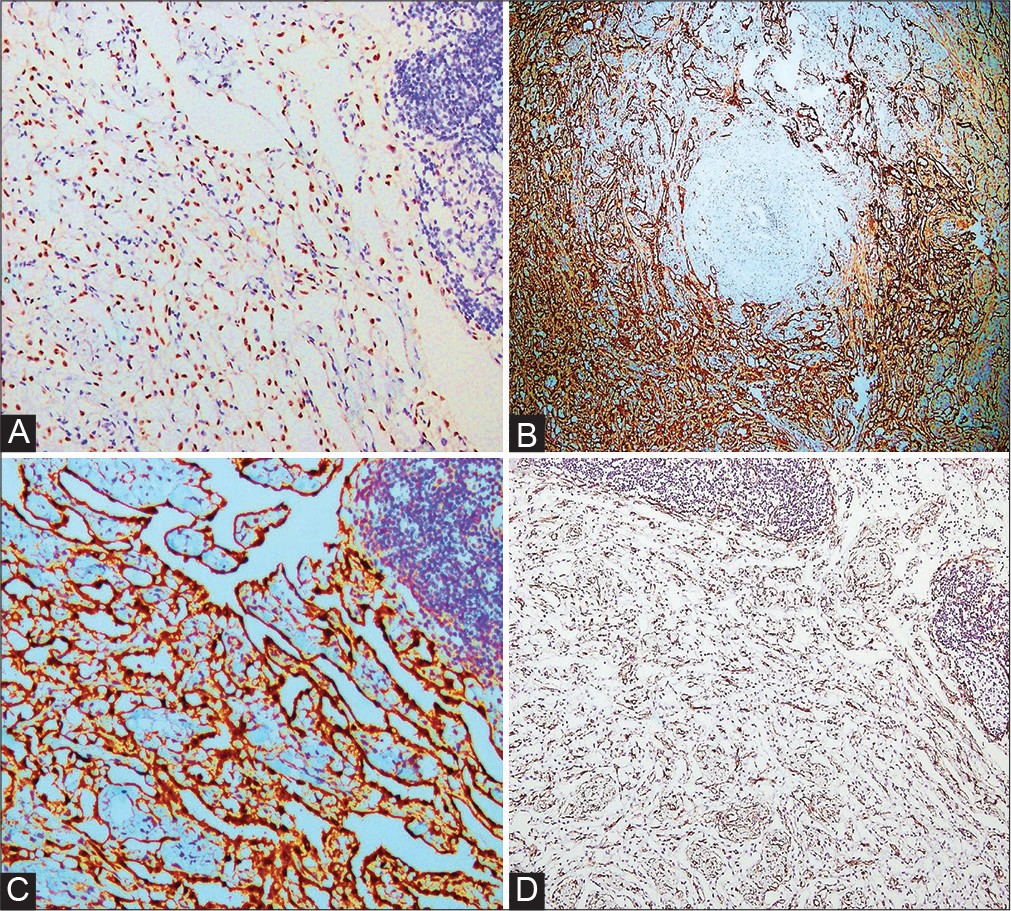
A 48-year-old female patient was brought to the hospital with complaints of pain in the right flank region and reduced urinary output for the past 1 month. Pain was intermittent, colicky, and nonradiating in nature. She also complained of intermittent high-grade fever since 2 months. Laboratory investigations revealed moderate anemia (hemoglobin 8.0 g/dL). Blood urea nitrogen was elevated (270 mg/dL) and serum creatinine was normal (0.8 mg/dL). The patient had been previously investigated outside and was diagnosed with right nonfunctioning kidney secondary to ureteric calculus along with an incidental right adnexal mass. Intraoperatively, a right complex tubo-ovarian cystic mass measuring 4 × 4 × 3 cm was identified. Laparoscopic right nephrectomy along with salpingo-oophorectomy was sent for histopathological examination. The histological features of the kidney were consistent with marked hydronephrosis with chronic pyelonephritis and mild uretritis. The right fallopian tube revealed a small nodular lesion with relatively circumscribed borders in the tubal wall (Figure 1A). The tumor was composed of closely packed slit-like elongated spaces, lined by bland looking plump ovoid cells which were intersecting between muscle bundles (Figure 1B, C). No cellular atypia or mitotic activity was noted (Figure 1D). The tubo-ovarian mass showed features of endometriotic cyst (Figure 2A, B). In addition, the tubal tumor revealed focal presence of few glandular spaces lined by bland ciliated columnar epithelium, surrounded by thin cuff of endometrial stromal cells, suggestive of intratumoral endometriosis (Figure 2C, D). On immunohistochemistry (IHC), the tumor cells were positive for Wilms tumor 1 (WT1) and calretinin (Figure 3A–C). The rich capillary network traversing in between the cellular component was highlighted by CD34 immunostain (Figure 3D). A final diagnosis of right tuboovarian endometriotic cyst along with incidental ipsilateral adenomatoid tumor with coexistent intratumoral endometriosis of the right fallopian tube was given. No further treatment was given to the patient. The patient is presently doing well in a 3-month follow-up with no signs of recurrence.
![(A) Low power view showing the presence of a well-circumscribed tumor in the fallopian tube wall (hematoxylin and eosin [HE] 40 ×). (B, C) Tumor composed of closely packed slit-like elongated spaces, intersecting between muscle bundles lined by bland looking plump ovoid cells (HE 100 ×). (D) Closely packed slit-like elongated spaces lined by bland looking plump ovoid cells (HE 200 ×).](/content/164/2024/16/1/img/JLP-16-120-g001.png)
- (A) Low power view showing the presence of a well-circumscribed tumor in the fallopian tube wall (hematoxylin and eosin [HE] 40 ×). (B, C) Tumor composed of closely packed slit-like elongated spaces, intersecting between muscle bundles lined by bland looking plump ovoid cells (HE 100 ×). (D) Closely packed slit-like elongated spaces lined by bland looking plump ovoid cells (HE 200 ×).
![(A) Surrounding tubal wall and part of ovary showing focal presence of endometriosis (arrows). (B) Low power view showing presence of endometriosis in the tubal wall (black arrow) and part of adenomatoid tumor (green arrow) (hematoxylin and eosin [HE] 40 ×). (C) Glandular spaces surrounded by thin cuff of endometrial stromal cells in the tubal tumor, suggestive of intratumoral endometriosis (HE 40 ×). (D) A glandular space lined by bland ciliated columnar epithelium, surrounded by thin cuff of endometrial stromal cells (HE 200 ×).](/content/164/2024/16/1/img/JLP-16-120-g002.png)
- (A) Surrounding tubal wall and part of ovary showing focal presence of endometriosis (arrows). (B) Low power view showing presence of endometriosis in the tubal wall (black arrow) and part of adenomatoid tumor (green arrow) (hematoxylin and eosin [HE] 40 ×). (C) Glandular spaces surrounded by thin cuff of endometrial stromal cells in the tubal tumor, suggestive of intratumoral endometriosis (HE 40 ×). (D) A glandular space lined by bland ciliated columnar epithelium, surrounded by thin cuff of endometrial stromal cells (HE 200 ×).

- Immunohistochemical stain. (A) Wilms tumor 1 (WT1): Nuclear positivity in the tumor cells (200 × ). (B) Calretinin: Positive in the tumor cells of adenomatoid tumor (endometriotic foci: negative). (C) Calretinin: Nuclear positivity in tumor cells (200 ×). (D) CD34 positivity highlighting the rich capillary network traversing in between the cellular component of tumor (40 ×).
DISCUSSION
Golden and Ash in 1945, first coined the term “adenomatoid tumor” and described it as a small, firm, asymptomatic intrascrotal mass, and considered to be of epithelial origin.[11] Ragins and Crane in accordance with their theory termed them as “adenoma/tubular adenoma.”[12] The older theory of an endothelial genesis has been discarded by most of the authors and has been correctly termed as adenomatoid tumor.[13] This terminology appears most suitable, due to the noncommittal origin of the lesion. Adenomatoid tumors are usually found in relation to the testicular tunica in the male, and to the uterus in the female.[14] Masson and Simard suggested the possible origin from the mesothelial lining by a process of dedifferentiation, most likely from misplaced cell rests.[15] This tumor, particularly lies in close approximation to the serous membranes and have shown clear-cut continuity of tumor cells with the lining mesothelial cells of the overlying serosa. In recent studies, the WT1 gene, which is involved in normal growth and differentiation of mesothelial tissue, has also been implicated in adenomatoid tumor development, further supporting its mesothelial origin.[16]
Adenomatoid tumors involving the fallopian tube is extremely rare and very few case reports have been published till date.[6–8] Lee et al described a case of positive BRCA1 mutation who underwent prophylactic total laparoscopic hysterectomy and bilateral salpingo-oophorectomy. Intraoperatively, a small nodular mass was identified in the fallopian tube which on histological examination showed closely packed small tubules with extensive psammoma bodies, thus mimicking serous carcinoma.[7] Similarly, in a recent report Zhang et al found an incidental tumor in the ampulla of the fallopian tube which morphologically showed presence of adenoids and lacunar-like structures of variable sizes and shapes in the hyperplastic smooth muscle tissue.[8]
Adenomatoid tumors display a spectrum of morphological patterns such as adenoid or tubular, glandular, angiomatoid, solid, cystic, or oncocytic forms. As a consequence, the differential diagnosis of these tumors is quite extensive, ranging from benign to malignant lesions of both epithelial and stromal origin. Ill-defined, infiltrative margins may lead to a mistaken diagnosis of carcinoma, including adenocarcinoma and mesothelioma; however, bland cytological features and absence of mitotic activity aids in their distinction. Tightly packed tubules and glands can mimic small solid nests on frozen section, a pattern commonly seen in lowgrade serous carcinoma. Another feature which might lead to a misdiagnosis of mucinous adenocarcinoma is the presence of vacuolated cells, thus imparting a signet ring morphology. Thus, it is imperative for pathologist to be aware of this benign entity and its varied morphological pattern which may lead to a potential misdiagnosis. IHC plays a pivotal role in diagnosing adenomatoid tumors and in differentiating it from other possible differentials. Adenomatoid tumor cells show strong positivity for cytokeratin, vimentin, calretinin, WT1, and D2–40. The absence of staining of epithelial markers like BerEP4/MOC31 and carcinoembryonic antigen is helpful in excluding carcinomas from the differential diagnosis, while negativity of vascular markers (CD34, factor excludes a diagnosis of hemangioma and lymphangioma. Although D2–40 has a high sensitivity and limited background staining, but this antigen is also used to identify the lining of lymphatic vessels (in cases of lymphangioma). Therefore, care must be taken not to rely on this stain completely to differentiate the two. Another lesion which may resemble adenomatoid tumor on histopathology is salpingitis isthmica nodosa (SIN) also called as diverticulosis of the fallopian tube. Here, the tubal epithelium outpouches into the muscular layer of the fallopian tube. The glands may be cystically dilated, and may communicate with the tubal lumen. The muscular layer shows hypertrophic and hyperplastic change. SIN stains negative for D2–40 and WT1 as opposed to adenomatoid tumor which is positive for mesothelial markers. Lipoleiomyoma and leiomyoma, another possible differential, shows positivity for S-100, smooth muscle actin (SMA), and only SMA, respectively, with negativity for epithelial and mesothelial markers. Considering the benign nature of this tumor, surgical excision is considered to be curative.
The other peculiar finding in the present case is the occurrence of intratumoral endometriosis within the adenomatoid tumor. The occurrence of coexistent endometriosis within any soft tissue tumor is extremely rare and only two cases have been published yet.[17,18] On extensive search, no such association has ever been reported with respect to adenomatoid tumor and this is the first ever case reported in literature.
CONCLUSIONS
Adenomatoid tumor is rarely encountered in surgical practice and is an incidental diagnosis on gross and microscopic examination. Due to rarity and varied histologic pattern, it may mimic other benign and malignant lesions, thus posing a diagnostic challenge to an unaware/inexperienced pathologist. Careful study of morphological features along with IHC markers will help in making the distinction and reaching an accurate diagnosis.
Guarantor of Submission
The corresponding author is the guarantor of submission.
Declaration of Patient Consent
The authors certify that they have obtained the appropriate consent from the patient. The patient has given his consent for the images and other clinical information to be reported in the journal. The parent understands that the name and initials will not be published, and due efforts have been made to conceal the same.
Ethical Standards
For this case report, formal consent from a local ethics committee is not required.
About The Author
Dr. Md Ali Osama is presently working as a Senior Resident in the Department of Pathology at Lady Hardinge Medical College, New Delhi. He has done his postgraduation (DNB) in Pathology from Sir Ganga Ram Hospital, New Delhi. His area of interest is in Oncopathology and Surgical Pathology. Besides, he is actively involved in research activities and has more than 33 publications in various national and international journals.
Conflict of Interest
None declared.
References
- Adenomatoid tumors of the female and male genital tracts: a clinicopathological and immunohistochemical study of 44 cases. Mod Pathol. 2009;22:1228-1235.
- [CrossRef] [PubMed] [Google Scholar]
- Adenomatoid tumor of the testis mimicking malignant testicular cancer on multiparametric ultrasound. Eur J Med Res. 2018;23:3.
- [CrossRef] [PubMed] [Google Scholar]
- Multicentric adenomatoid tumors involving uterus, ovary, and appendix. J Obstet Gynaecol Res. 2003;29:234-238.
- [CrossRef] [PubMed] [Google Scholar]
- Adenomatoid tumor of the pancreas: a case report with comparison of histology and aspiration cytology. Mod Pathol. 2003;16:613-617.
- [CrossRef] [PubMed] [Google Scholar]
- Cystic adenomatoid tumor of the mediastinum. Am J Surg Pathol. 2004;28:132-138.
- [CrossRef] [PubMed] [Google Scholar]
- Adenomatoid tumor in the fallopian tube a rare case. Int J Pathol Clin Res. 2019;5:96.
- [CrossRef] [Google Scholar]
- An unusual adenomatoid tumor of fimbria with pronounced psammoma bodies in a BRCA positive patient as a pitfall for carcinoma on frozen section. Case Rep Pathol. 2018;2018:8148147.
- [CrossRef] [PubMed] [Google Scholar]
- Adenomatoid tumor of the fallopian tube. Asian J Surg. 2022;45:754-755.
- [CrossRef] [PubMed] [Google Scholar]
- Adenomatoid tumor of the uterus; report of a rare incidentaloma. Int J Reprod Contracept Obstet Gynecol. 2014;3:769-771.
- [CrossRef] [Google Scholar]
- Tumors of the broad ligament: what and when to suspect such rare location. Radiol Bras. 2020;53:349-355.
- [CrossRef] [PubMed] [Google Scholar]
- The histogenesis of the adenomatoid tumor of the genital tract. Cancer. 1958;11:337-350.
- [CrossRef] [PubMed] [Google Scholar]
- Adenomatoid tumor: a review of pathology with focus on unusual presentations and sites, histogenesis, differential diagnosis, and molecular and clinical aspects with a historic overview of its description. Adv Anat Pathol. 2020;27:394-407.
- [CrossRef] [PubMed] [Google Scholar]
- Adenomatoid tumors of the female and male genital tracts express WT1. Int J Gynecol Pathol. 2004;23:123-128.
- [CrossRef] [PubMed] [Google Scholar]
- Intratumoral endometriosis in renal angiomyolipomas associated to medium vessel necrotizing vasculitis in a patient with Wünderlich syndrome. A rare finding [in Spanish] Rev Esp Patol. 2019;52:202-204.
- [CrossRef] [PubMed] [Google Scholar]
- Gastrointestinal stromal tumour with intratumoural endometriosis. J Clin Diagn Res. 2021;15:EJ01-EJ02.
- [CrossRef] [Google Scholar]


