
Translate this page into:
Cholecystitis Associated with Heterotopic Pancreas, Pseudopyloric Metaplasia, and Adenomyomatous Hyperplasia: A Rare Combination
Address for correspondence: Dr. Navjot Kaur, E-mail: drjagjitschahal@yahoo.co.in
This is an open access article distributed under the terms of the Creative Commons Attribution NonCommercial ShareAlike 3.0 License, which allows others to remix, tweak, and build upon the work non commercially, as long as the author is credited and the new creations are licensed under the identical terms.
This article was originally published by Medknow Publications & Media Pvt Ltd and was migrated to Scientific Scholar after the change of Publisher.
Abstract
Heterotopic pancreatic tissue in the gall bladder is an uncommon incidental finding in most cases. We hereby describe the case of a 45-year-old woman who presented with symptoms of acalculous cholecystitis. Pathological examination detected heterotopic pancreatic tissue, pseudopyloric metaplasia, and adenomyomatous hyperplasia in the gall bladder. This is a rare combination of three entities which is being reported for the first time. This case emphasizes that heterotopic pancreas might be the causative factor for cholecystitis.
Keywords
Adenomyomatous hyperplasia
chronic cholecystitis
gall bladder
heterotopic pancreas
pseudopyloric metaplasia

INTRODUCTION
Heterotopic pancreas (HP) is defined as the presence of pancreatic tissue lying outside its normal location and lacking anatomical or vascular continuity with the pancreas proper.[1] In 85–90% of reported cases, HP has been found in stomach, duodenum, upper jejunum, whereas its presence in the gallbladder is very rare.[12] Despite its congenital origin, pancreatic heterotopia is usually diagnosed during adult life.[34] As it is asymptomatic most of the time, a definitive diagnosis is made on histopathological examination in a gall bladder, removed for other indications.[35] We report a case of HP of the gallbladder along with pseudopyloric metaplasia and segmental adenomyomatous hyperplasia in a 45-year-old woman. Up to the present study, about 31 cases of HP in the gall bladder have been reported,[5] but this is the first of its kind which has two other histopathological findings associated with it.
CASE REPORT
A 45-year-old woman presented to our hospital with 2 months history of the right upper quadrant abdominal pain along with nausea and vomiting. Her history was unremarkable. All her vitals were stable. Routine blood investigations including renal and liver function tests revealed no abnormality. On physical examination, there was tenderness in the right upper quadrant. Abdominal ultrasound showed no abnormality. Based on the diagnosis of cholecystitis, cholecystectomy was carried out.
On gross examination, the gall bladder measured 7 cm in length and 2.5 cm in circumference with a wall thickness ranging from 0.2 to 0.4 cm. The serosa was unremarkable. Cut section revealed velvety green mucosa.
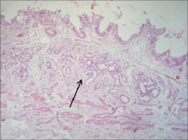
On microscopic examination, there was a well-circumscribed rest of heterotopic pancreatic tissue on the serosal aspect of the gall bladder wall, composed of lobules of exocrine pancreatic acini and an occasional duct. Islets of Langerhans were not seen. Mucosal layer adjacent to the pancreatic tissue showed adenomyomatous hyperplasia [Figure 1]. A focus elsewhere showed pseudopyloric metaplasia apart from histological features of chronic cholecystitis [Figure 2].

- Photomicrograph of gall bladder showing a well-circumscribed rest of heterotopic pancreas (arrowhead) and adjacent mucosa showing adenomyomatous hyperplasia (arrow) (H and E, ×40). Inset shows lobules of pancreatic acini and an occasional duct (H and E, ×400)
- Photomicrograph showing pseudopyloric metaplasia in a separate focus along with features of chronic cholecystitis (H and E, ×40)
The diagnosis was thereby established as chronic cholecystitis with heterotopic pancreatic tissue, pseudopyloric metaplasia, and segmental adenomyomatous hyperplasia of the gall bladder.
The patient's postoperative course was uneventful and was discharged after 5 days without any complications. After a follow-up of 2 months, the patient was asymptomatic and had recovered fully.
DISCUSSION
Although HP is the second most prevalent pancreatic anomaly, the incidence in gastrointestinal tract is estimated to be from 0.55% to 13.7% on autopsy and 0.2% on laparotomy.[6] Despite the frequent occurrence of HP in the stomach, duodenum, and upper jejunum, the gallbladder localization is extremely rare.[7]
Heterotopic tissue is usually located in the neck or fundus of the gall bladder, varies in size from 0.1 to 1.0 cm, and may exhibit several patterns, ranging from intramural to exophytic to polypoidal lesions.[36] As there is no submucosal layer in the gall bladder, HP is usually seen in the muscularis. Microscopic examination shows a varying degree of excretory ducts, exocrine glands, and islets of Langerhans.[8] Microscopically, HP has been classified into three types by von Heinrich - Type 1: Ectopic tissue with acini, ducts, and islets of Langerhans; Type 2: Ectopic tissue containing only a few acini and ducts, with absence of endocrine elements - incomplete arrangement; Type 3: Ectopic tissue with only proliferating excretory ducts and absence of exocrine acini and endocrine elements.[9] Our case was considered to be Type 2, based on the Heinrich classification.
A recent theory suggested that abnormalities in the notch signaling system, a main factor for lesion - appropriate pancreatic differentiation in the development of the foregut endoderm, lead to the development of heterotopic pancreatic tissue. However, there is no accepted theory that explains the exact origin of an HP.[5]
CONCLUSION
Chronic cholecystitis with heterotopic pancreatic tissue, pseudopyloric metaplasia, and adenomyomatous hyperplasia of the gall bladder is rarely encountered. HP of the gall bladder itself is a very rare condition which is usually diagnosed incidentally, but may cause symptoms of benign gall bladder disease without a definitive lesion on radiology. Awareness of this under-reported condition may help in its recognition and this, in turn, may shed more light on its clinical significance. In our case, we could not discriminate whether the patient's symptoms were caused by HP or by segmental adenomyomatous hyperplasia of the gall bladder.
Financial support and sponsorship
Nil.
Conflicts of interest
There are no conflicts of interest.
REFERENCES
- The clinical significance of heterotopic pancreas in the gastrointestinal tract. Br J Surg. 1981;68:384-7.
- [Google Scholar]
- The fate of heterotopic pancreatic tissue. A study of 212 cases. Arch Surg. 1974;109:762-5.
- [Google Scholar]
- Heterotopic pancreas in gall bladder associated with chronic cholecystolithiasis. Int J Appl Basic Med Res. 2012;2:142-3.
- [Google Scholar]
- Heterotopic pancreatic tissue located in the gallbladder wall. A case report. JOP. 2011;12:152-4.
- [Google Scholar]
- Heterotopic pancreas of the gallbladder associated with segmental adenomyomatosis of the gallbladder. J Korean Surg Soc. 2013;84:309-11.
- [Google Scholar]
- An unusual cause of hydropic gallbladder and biliary colic – Heterotopic pancreatic tissue in the cystic duct: Report of a case and review of the literature. Surg Today. 1993;23:532-4.
- [Google Scholar]
- An unusual cause of cholecystitis: Heterotopic pancreatic tissue in the gallbladder. World J Gastroenterol. 2007;13:313-5.
- [Google Scholar]
- Pancreatic heterotopia: A reappraisal and clinicopathologic analysis of 32 cases. South Med J. 1988;81:1264-75.
- [Google Scholar]
- Case of intra-abdominal endocrine tumor possibly arising from an ectopic pancreas. J Nippon Med Sch. 2007;74:168-72.
- [Google Scholar]


