
Translate this page into:
Exflagellation of Plasmodium vivax in peripheral blood: An uncommon finding and its significance
Address for correspondence: Dr. Gaurav Chhabra, Department of Pathology and Laboratory Medicine, AIIMS, Bhubaneswar, Odisha, India. E-mail: gauravchhabra2001@gmail.com
-
Received: ,
Accepted: ,
This is an open access journal, and articles are distributed under the terms of the Creative Commons Attribution-NonCommercial-ShareAlike 4.0 License, which allows others to remix, tweak, and build upon the work non-commercially, as long as appropriate credit is given and the new creations are licensed under the identical terms.
This article was originally published by Wolters Kluwer - Medknow and was migrated to Scientific Scholar after the change of Publisher.
Abstract
Malaria continues to be a major public health problem. The life cycle of malaria is completed in two hosts Anopheles mosquito – definitive host and humans – the intermediate host. Exflagellation of microgametocyes in the life cycle of Plasmodium vivax occurs in mosquitoes and is rarely seen in human peripheral blood. Less than 15 occurrences of exflagellated microgametocyte of Plasmodium species have been reported to date. The appearance of exflagellated microgametes in human blood may pose a diagnostic dilemma due to its resemblance with other hemoparasites such as Borrelia and Trypanosoma.
Keywords
Exflagellation
hemoparasites
malaria
microgametocyte
Plasmodium

Introduction
Malaria is a major public health issue, particularly in Africa and Southeast Asia. It is caused due to infection with Plasmodium protozoa. The life cycle of Plasmodium species is complex and multistage, with sexual stage occurring in infected female Anopheles mosquito (definitive host) and asexual stage occurring in humans (intermediate host). All the asexual stages of Plasmodium vivax such as ring forms, late trophozoites, schizonts, and gametocytes are observed commonly in human blood whereas, sexual stages such as exflagellation of microgametocyte and microgametes are rarely seen in humans. Less than 15 occurrences of exflagellated microgametocyte of Plasmodium spp. have been reported to date. Herein, we report a case of P. vivax infection with different stages of exflagellation and numerous exflagellated microgametes in addition to the ring forms of P. vivax in peripheral blood of a patient with high-grade fever and its significance in the clinical laboratory diagnosis.
Case Report
A 70-year-old female patient presented in the emergency department with high-grade fever for the last 2 days. The patient's blood sample was sent to the laboratory in ethylenediaminetetraacetic acid (EDTA) vacutainer for complete hemogram analysis. On receipt, in the laboratory, the sample was run on XT4000i (Sysmex, Kobe, Japan) automated hematology analyzer. The hemogram analysis revealed total leukocyte count of 11.43/μl with a system generated flag of abnormal white blood cell scattergram, and no differential count was reported by the analyzer. The scatter plot revealed no clear-cut separation of neutrophil and eosinophil population in WDF channel on SFL (Side Fluorescence) versus SSC (Side Scatter) plot, indicating the presence of hemoparasite [Figure 1]. Since no differential was given with an abnormal flag, a peripheral smear (PS) review was necessitated. The PS was made and stained with Leishman stain and examined under the microscope which revealed numerous ring forms, trophozoites and schizonts of P. vivax [Figure 2a]. In addition, multiple clusters as well as singly scattered thin, long, filamentous flagellae-like structures were observed outside the red blood cells which were approximately around 10–15 μ in length and having oval-shaped nucleus. The careful morphological examination made us suspect the presence of exflagellated microgametes of P. vivax. On further examination, different stages of exflagellation of microgametocyes with the initial stage of 6–8 flagellated microgametes arising out of microgametocyte [Figure 2b-2e] followed by detached single exflagellated microgametes [Figure 2f] were found. To corroborate our finding, we made a fresh smear from finger-prick blood in which the exflagellated forms were absent.
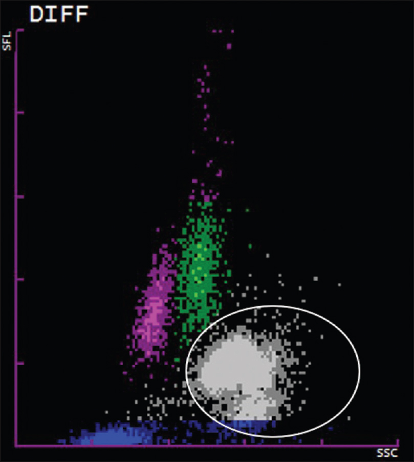
- White blood cell scatter plot showing abnormal white blood cell scattergram (encircled population)
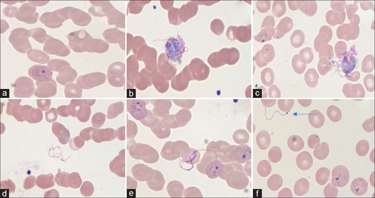
- (a) Trophozoite of Plasmodium vivax (×1000; Leishman). (b and c) Exflagellation of microgametes from microgametocytes (×1000; Leishman). (d-f) Exflagellated microgametes (×1000; Leishman)
Discussion
Exflagellation of Plasmodium microgametocytes occurs in mosquito and its appearance in human peripheral blood is an extremely rare phenomenon. In 1897, MacCallum[1] first observed and reported this event in a patient with Plasmodium falciparum infection. On extensive search of English literature, Less than 15 occurrences[2345678] have been reported in the form of case reports. To the best of our knowledge, this is the first case reported from India in which various stages of exflagellation from microgametocytes with resultant formation of microgamete have been found in human blood.
Microgametogenesis and exflagellation in vitro is dependent on many factors such as rise in pH, pCO2, bicarbonate levels, and fall in temperature below that of the vertebrate host and anticoagulant.[91011] Mosquito exflagellation factor (MEF), a heat-stable molecule derived from mosquito's head and gut is considered as a most potent factor for in vitro exflagellation.[12] The activity of MEF is dependent on the pH and bicarbonate level. The pH of human blood is around 7.35–7.45, which is lower than pH of mosquito gut, which inhibits the microgametogenesis and exflagellation. It is hypothesized that a change in pH in the laboratory may be triggered, when the blood comes in contact with the atmospheric environment resulting a fall in CO2, ultimately raising the pH. This scenario mimics the change, the gametocytes are exposed to when they reach the gut of the mosquito following an infective meal, thus favoring exflagellation.[7] In our case, exflagellation was not observed in smears prepared immediately through finger prick, thus confirming the fact that exflagellation was induced when the blood got exposed to atmospheric air for a longer duration, resulting in pH change.
Other reports suggest that phosphodiesterase inhibitors and the use of caffeine can induce this phenomenon in vitro; however, the mechanism is not clear.[1213] Solarte et al. suggested that the use of certain anticoagulant-like heparin induces exflagellation by causing a pH change, while EDTA inhibits it by preventing pH change, consumption of Ca2+, Mn2+, and Mg2+, which further prevents the activation of enzymes required for exflagellation.[14] EDTA-induced prevention of flagellation is questionable, as majority of cases published reported exflagellation in the blood preserved with EDTA as seen in our case.
Due to its rarity, the presence of exflagellated microgametes in peripheral blood may pose a diagnostic dilemma, particularly for an inexperienced observer because of its resemblance with other hemoparasites such as Borrelia, Microfilaria, or Trypanosoma or it may be overlooked as a staining artifact. However, with careful morphological examination, these can be differentiated as the exflagellated form of Plasmodium occur as thin filamentous structure measuring approximately 10–15 μ in length with an oval-shaped dark blue nucleus. In contrast, Borrelia is 5–20 μ in length with a spiral shape and lack nuclei and Trypanosoma has an undulating membrane, kinetoplast, and a nucleus.[34]
Conclusion
The presence of exflagellated microgametes in human peripheral blood is a rare phenomenon and this is possibly the third reported case from India, an endemic zone for malaria. Lack of knowledge of this poses a diagnostic challenge because of its resemblance with other hemoparasites. Careful morphological examination can help in correctly diagnosing and differentiating these forms from other hemoparasites.
Declaration of patient consent
The authors certify that they have obtained all appropriate patient consent forms. In the form the patient(s) has/have given his/her/their consent for his/her/their images and other clinical information to be reported in the journal. The patients understand that their names and initials will not be published and due efforts will be made to conceal their identity, but anonymity cannot be guaranteed.
Financial support and sponsorship
Nil.
Conflicts of interest
There are no conflicts of interest.
References
- Exflagellation of malarial parasites in human peripheral blood. J Clin Microbiol. 1981;13:236-7.
- [Google Scholar]
- Exflagellated microgametes of Plasmodium vivax in human peripheral blood: A case report and review of the literature. Indian J Pathol Microbiol. 2009;52:252-4.
- [Google Scholar]
- Exflagellated microgametes of Plasmodium vivax in human peripheral blood: An uncommon feature of malaria. Indian J Hematol Blood Transfus. 2011;27:104-6.
- [Google Scholar]
- Images in clinical medicine. In vitro exflagellation of Plasmodium vivax. N Engl J Med. 2016;375:e27.
- [Google Scholar]
- Plasmodium falciparum exflagellation in a patient with ovalocytosis. Am J Med Sci. 2017;354:68.
- [Google Scholar]
- Occurrence of exflagellation and microgametes in peripheral blood of a patient with malaria. J Infect Dis. 1982;146:448.
- [Google Scholar]
- Exflagellation of microgametocytes in Plasmodium vivax malaria: A diagnostic conundrum. Med Princ Pract. 2004;13:298-300.
- [Google Scholar]
- Control of gamete formation (exflagellation) in malaria parasites. Science. 1977;195:407-9.
- [Google Scholar]
- The roles of temperature, pH and mosquito factors as triggers of male and female gametogenesis of Plasmodium berghei in vitro. Parasitology. 1997;115(Pt 1):1-7.
- [Google Scholar]
- Plasmodium gallinaceum: Exflagellation stimulated by a mosquito factor. Exp Parasitol. 1979;48:75-80.
- [Google Scholar]
- Plasmodium gallinaceum: Induction of male gametocyte exflagellation by phosphodiesterase inhibitors. Exp Parasitol. 1978;44:239-42.
- [Google Scholar]
- Effects of anticoagulants on Plasmodium vivax oocyst development in Anopheles albimanus mosquitoes. Am J Trop Med Hyg. 2007;77:242-5.
- [Google Scholar]


